1U9V

Crystal Structure of the Cysteine Protease Human Cathepsin K in Complex with the Covalent Inhibitor NVP-ABE854


Experimental Data Snapshot

  • Method: X-RAY DIFFRACTION
  • Resolution: 2.20 Å
  • R-Value Free: 0.231 
  • R-Value Work: 0.170 
  • R-Value Observed: 0.170 

wwPDB Validation   3D Report Full Report


Ligand Structure Quality Assessment 


This is version 1.4 of the entry. See complete history


Literature

Novel purine nitrile derived inhibitors of the cysteine protease cathepsin K

Altmann, E.Cowan-Jacob, S.W.Missbach, M.

(2004) J Med Chem 47: 5833-5836

  • DOI: https://doi.org/10.1021/jm0493111
  • Primary Citation of Related Structures:  
    1U9V, 1U9W, 1U9X

  • PubMed Abstract: 

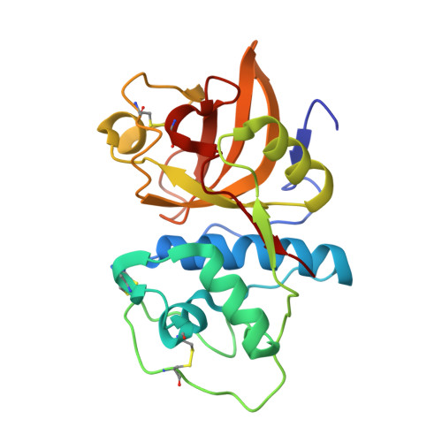
    Starting from the high-throughput screening hit 1a, novel cathepsin K inhibitors have been developed based on a purine scaffold. High-resolution X-ray structures of several derivatives have revealed the binding mode of these unique cysteine protease inhibitors.


  • Organizational Affiliation

    Novartis Institutes for BioMedical Research, CH-4002 Basel, Switzerland. eva.altmann@pharma.novartis.com


Macromolecules
Find similar proteins by:  (by identity cutoff)  |  3D Structure
Entity ID: 1
MoleculeChains Sequence LengthOrganismDetailsImage
Cathepsin K217Homo sapiensMutation(s): 0 
EC: 3.4.22.38
UniProt & NIH Common Fund Data Resources
Find proteins for P43235 (Homo sapiens)
Explore P43235 
Go to UniProtKB:  P43235
PHAROS:  P43235
GTEx:  ENSG00000143387 
Entity Groups  
Sequence Clusters30% Identity50% Identity70% Identity90% Identity95% Identity100% Identity
UniProt GroupP43235
Sequence Annotations
Expand
  • Reference Sequence
Small Molecules
Ligands 1 Unique
IDChains Name / Formula / InChI Key2D Diagram3D Interactions
IHE
Query on IHE

Download Ideal Coordinates CCD File 
B [auth A]6-(CYCLOHEXYLAMINO)-9-[2-(4-METHYLPIPERAZIN-1-YL)-ETHYL]-9H-PURINE-2-CARBONITRILE
C19 H28 N8
OGODDSLNRULSMM-UHFFFAOYSA-N
Binding Affinity Annotations 
IDSourceBinding Affinity
IHE PDBBind:  1U9V IC50: 6 (nM) from 1 assay(s)
BindingDB:  1U9V IC50: 6 (nM) from 1 assay(s)
Experimental Data & Validation

Experimental Data

  • Method: X-RAY DIFFRACTION
  • Resolution: 2.20 Å
  • R-Value Free: 0.231 
  • R-Value Work: 0.170 
  • R-Value Observed: 0.170 
  • Space Group: P 43 21 2
Unit Cell:
Length ( Å )Angle ( ˚ )
a = 63.2α = 90
b = 63.2β = 90
c = 113.8γ = 90
Software Package:
Software NamePurpose
XDSdata scaling
AUTOMARdata reduction
AMoREphasing
X-PLORrefinement
XDSdata reduction

Structure Validation

View Full Validation Report


Ligand Structure Quality Assessment 


Entry History 

Deposition Data

Revision History  (Full details and data files)

  • Version 1.0: 2005-08-11
    Type: Initial release
  • Version 1.1: 2008-04-30
    Changes: Version format compliance
  • Version 1.2: 2011-07-13
    Changes: Version format compliance
  • Version 1.3: 2017-10-11
    Changes: Refinement description
  • Version 1.4: 2023-10-25
    Changes: Data collection, Database references, Derived calculations, Refinement description