1U0B

Crystal structure of cysteinyl-tRNA synthetase binary complex with tRNACys


Experimental Data Snapshot

  • Method: X-RAY DIFFRACTION
  • Resolution: 2.30 Å
  • R-Value Free: 0.277 
  • R-Value Work: 0.233 
  • R-Value Observed: 0.250 

wwPDB Validation   3D Report Full Report


This is version 1.3 of the entry. See complete history


Literature

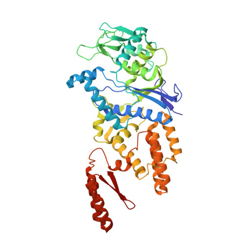
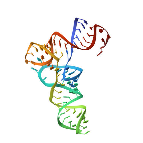
Shape-selective RNA recognition by cysteinyl-tRNA synthetase

Hauenstein, S.Zhang, C.M.Hou, Y.M.Perona, J.J.

(2004) Nat Struct Mol Biol 11: 1134-1141

  • DOI: https://doi.org/10.1038/nsmb849
  • Primary Citation of Related Structures:  
    1U0B

  • PubMed Abstract: 

    The crystal structure of Escherichia coli cysteinyl-tRNA synthetase (CysRS) bound to tRNA(Cys) at a resolution of 2.3 A reveals base-specific and shape-selective interactions across an extensive protein-RNA recognition interface. The complex contains a mixed alpha/beta C-terminal domain, which is disordered in the unliganded enzyme. This domain makes specific hydrogen bonding interactions with all three bases of the GCA anticodon. The tRNA anticodon stem is bent sharply toward the enzyme as compared with its conformation when bound to elongation factor Tu, providing an essential basis for shape-selective recognition. The CysRS structure also reveals interactions of conserved enzyme groups with the sugar-phosphate backbone in the D loop, adjacent to an unusual G15.G48 tertiary base pair previously implicated in tRNA aminoacylation. A combined mutational analysis of enzyme and tRNA groups at G15.G48 supports the notion that contacts between CysRS and the sugar-phosphate backbone contribute to recognition by indirect readout.


  • Organizational Affiliation

    Department of Chemistry and Biochemistry, University of California at Santa Barbara, Santa Barbara, California 93106-9510, USA.


Macromolecules

Find similar proteins by:  (by identity cutoff)  |  3D Structure
Entity ID: 2
MoleculeChains Sequence LengthOrganismDetailsImage
Cysteine--tRNA ligase461Escherichia coliMutation(s): 0 
Gene Names: cysS
EC: 6.1.1.16
UniProt
Find proteins for P21888 (Escherichia coli (strain K12))
Explore P21888 
Go to UniProtKB:  P21888
Entity Groups  
Sequence Clusters30% Identity50% Identity70% Identity90% Identity95% Identity100% Identity
UniProt GroupP21888
Sequence Annotations
Expand
  • Reference Sequence
Find similar nucleic acids by:  (by identity cutoff)  |  3D Structure
Entity ID: 1
MoleculeChains LengthOrganismImage
cysteinyl tRNA74Escherichia coli
Sequence Annotations
Expand
  • Reference Sequence
Small Molecules
Ligands 1 Unique
IDChains Name / Formula / InChI Key2D Diagram3D Interactions
ZN
Query on ZN

Download Ideal Coordinates CCD File 
C [auth B]ZINC ION
Zn
PTFCDOFLOPIGGS-UHFFFAOYSA-N
Experimental Data & Validation

Experimental Data

  • Method: X-RAY DIFFRACTION
  • Resolution: 2.30 Å
  • R-Value Free: 0.277 
  • R-Value Work: 0.233 
  • R-Value Observed: 0.250 
  • Space Group: P 41 21 2
Unit Cell:
Length ( Å )Angle ( ˚ )
a = 129.49α = 90
b = 129.49β = 90
c = 91.02γ = 90
Software Package:
Software NamePurpose
DENZOdata reduction
SCALEPACKdata scaling
CNSrefinement
CNSphasing

Structure Validation

View Full Validation Report


Entry History 

Deposition Data

Revision History  (Full details and data files)

  • Version 1.0: 2004-11-23
    Type: Initial release
  • Version 1.1: 2008-04-30
    Changes: Version format compliance
  • Version 1.2: 2011-07-13
    Changes: Version format compliance
  • Version 1.3: 2023-08-23
    Changes: Data collection, Database references, Derived calculations, Refinement description, Source and taxonomy, Structure summary