1TXY

E. coli PriB


Experimental Data Snapshot

  • Method: X-RAY DIFFRACTION
  • Resolution: 2.00 Å
  • R-Value Free: 0.285 
  • R-Value Work: 0.259 
  • R-Value Observed: 0.260 

wwPDB Validation   3D Report Full Report


This is version 1.2 of the entry. See complete history


Literature

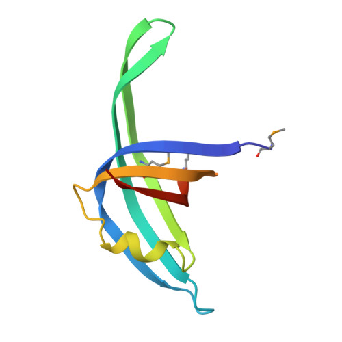
Crystal structure of PriB, a component of the Escherichia coli replication restart primosome

Lopper, M.Holton, J.M.Keck, J.L.

(2004) Structure 12: 1967-1975

  • DOI: https://doi.org/10.1016/j.str.2004.09.004
  • Primary Citation of Related Structures:  
    1TXY

  • PubMed Abstract: 

    Maintenance of genome stability following DNA damage requires origin-independent reinitiation of DNA replication at repaired replication forks. In E. coli, PriA, PriB, PriC, and DnaT play critical roles in recognizing repaired replication forks and reloading the replisome onto the template to reinitiate DNA replication. Here, we report the 2.0 A resolution crystal structure of E. coli PriB, revealing a dimer that consists of a single structural domain formed by two oligonucleotide/oligosaccharide binding (OB) folds. Structural similarity of PriB to single-stranded DNA binding proteins reveals insights into its mechanisms of DNA binding. The structure further establishes a putative protein interaction surface that may contribute to the role of PriB in primosome assembly by facilitating interactions with PriA and DnaT. This is the first high-resolution structure of a protein involved in oriC-independent replisome loading and provides unique insight into mechanisms of replication restart in E. coli.


  • Organizational Affiliation

    Department of Biomolecular Chemistry, University of Wisconsin Medical School, 550 Medical Sciences Center, 1300 University Avenue, Madison, WI 53706, USA.


Macromolecules
Find similar proteins by:  (by identity cutoff)  |  3D Structure
Entity ID: 1
MoleculeChains Sequence LengthOrganismDetailsImage
Primosomal replication protein n
A, B
104Escherichia coliMutation(s): 2 
Gene Names: PRIBB4201
UniProt
Find proteins for P07013 (Escherichia coli (strain K12))
Explore P07013 
Go to UniProtKB:  P07013
Entity Groups  
Sequence Clusters30% Identity50% Identity70% Identity90% Identity95% Identity100% Identity
UniProt GroupP07013
Sequence Annotations
Expand
  • Reference Sequence
Small Molecules
Modified Residues  1 Unique
IDChains TypeFormula2D DiagramParent
MSE
Query on MSE
A, B
L-PEPTIDE LINKINGC5 H11 N O2 SeMET
Experimental Data & Validation

Experimental Data

  • Method: X-RAY DIFFRACTION
  • Resolution: 2.00 Å
  • R-Value Free: 0.285 
  • R-Value Work: 0.259 
  • R-Value Observed: 0.260 
  • Space Group: P 21 21 21
Unit Cell:
Length ( Å )Angle ( ˚ )
a = 49.902α = 90
b = 60.294β = 90
c = 66.304γ = 90
Software Package:
Software NamePurpose
REFMACrefinement
MOSFLMdata reduction
CCP4data scaling
SOLVEphasing

Structure Validation

View Full Validation Report


Entry History 

Deposition Data

Revision History  (Full details and data files)

  • Version 1.0: 2004-11-16
    Type: Initial release
  • Version 1.1: 2008-04-30
    Changes: Version format compliance
  • Version 1.2: 2011-07-13
    Changes: Advisory, Refinement description, Version format compliance