Solution structure of the TR1C fragment of skeletal muscle troponin-C.
Findlay, W.A., Sonnichsen, F.D., Sykes, B.D.(1994) J Biol Chem 269: 6773-6778
- PubMed: 8120037
- DOI: https://doi.org/10.2210/pdb1trf/pdb
- Primary Citation of Related Structures:
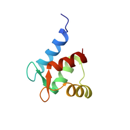
1TRF - PubMed Abstract:
Residues 12-87 (TR1C fragment) of turkey skeletal muscle troponin-C comprises two helix-loop-helix calcium-binding motifs which are the regulatory calcium-binding sites in the N-terminal domain of the protein. We have used the combined distance geometry-simulated annealing protocol DGII (Havel, T. F. (1991) Prog. Biophys. Mol. Biol. 56, 43-78) to determine the structure of this 76-residue polypeptide in solution from 475 1H NMR-derived distance restraints. The nuclear Overhauser enhancement-derived distance constraints used in the DGII protocol were supplemented by introducing generic hydrogen bond distance restraints for slowly exchanging amide hydrogens in regular secondary structure elements, by restricting the available phi angle space to -180 degrees to 0 degrees for all residues except glycines, and by tailoring the distance boundaries used for quantitating the nuclear Overhauser enhancement intensities to correspond to characteristic distances found in helices. This improved the geometry of the four helices in the resulting structures. The relative positions of helices A and B which flank calcium-binding loop 1, helix D which follows calcium-binding loop 2, and the beta-sheet between the two calcium-binding loops were well defined and had an overall root-mean-square deviation for 20 converged structures of 1.4 +/- 0.2 A for backbone atoms. The structure and relative orientations of these regions are very similar to these of the corresponding regions of the protein in the crystal structure of intact turkey skeletal troponin C (Herzberg, O., and James, M. N. G. (1988) Nature 313, 653-659). The structure of helix C was well defined, but its relative position to the other helices was not defined. It occupied a range of positions in the set of 20 DGII structures, the average of which was quite similar to the orientation of helix C in the x-ray structure. The overall structure of the apo regulatory domain of troponin-C is therefore not affected by the loss of the N-helix, or the low pH conditions used for the x-ray structure, but may be more flexible in regions known to be involved in contacts with other skeletal muscle regulatory proteins.
Organizational Affiliation:
Department of Biochemistry, University of Alberta, Edmonton, Canada.