1TMC

THE THREE-DIMENSIONAL STRUCTURE OF A CLASS I MAJOR HISTOCOMPATIBILITY COMPLEX MOLECULE MISSING THE ALPHA3 DOMAIN OF THE HEAVY CHAIN


Experimental Data Snapshot

  • Method: X-RAY DIFFRACTION
  • Resolution: 2.30 Å
  • R-Value Work: 0.207 
  • R-Value Observed: 0.207 

wwPDB Validation   3D Report Full Report


This is version 1.2 of the entry. See complete history


Literature

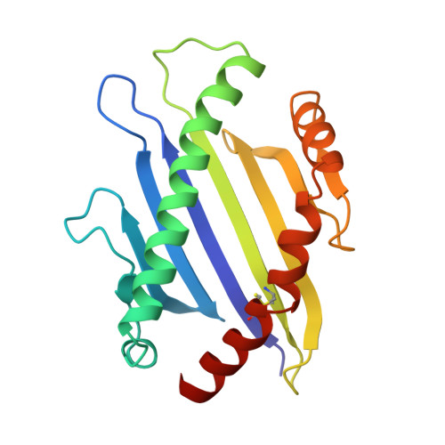
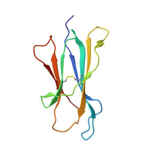
The three-dimensional structure of a class I major histocompatibility complex molecule missing the alpha 3 domain of the heavy chain.

Collins, E.J.Garboczi, D.N.Karpusas, M.N.Wiley, D.C.

(1995) Proc Natl Acad Sci U S A 92: 1218-1221

  • DOI: https://doi.org/10.1073/pnas.92.4.1218
  • Primary Citation of Related Structures:  
    1TMC

  • PubMed Abstract: 

    Class I major histocompatibility complex (MHC) molecules are ternary complexes of the soluble serum protein beta 2-microglobulin, MHC heavy chain, and bound peptide. The first two domains (alpha 1, alpha 2) of the heavy chain create the peptide binding cleft and the surface that contacts the T-cell receptor. The third domain (alpha 3) associates with the T-cell co-receptor, CD8, during T-cell recognition. Here we describe the x-ray crystal structure of a human class I MHC molecule, HLA-Aw68, from which the alpha 3 domain has been proteolytically removed. The resulting molecule shows no gross morphological changes compared to the intact protein. A decameric peptide complexed with the intact HLA-Aw68 is seen to bind to the proteolized molecule in the conventional manner, demonstrating that the alpha 3 domain is not required for the structural integrity of the molecule or for peptide binding.


  • Organizational Affiliation

    Department of Molecular and Cellular Biology, Howard Hughes Medical Institute, Harvard University, Cambridge, MA 02138.


Macromolecules
Find similar proteins by:  (by identity cutoff)  |  3D Structure
Entity ID: 1
MoleculeChains Sequence LengthOrganismDetailsImage
CLASS I HISTOCOMPATIBILITY ANTIGEN (HLA-AW68)175Homo sapiensMutation(s): 0 
Gene Names: BETA-2-MICROGLOBULIN
UniProt & NIH Common Fund Data Resources
Find proteins for P04439 (Homo sapiens)
Explore P04439 
Go to UniProtKB:  P04439
PHAROS:  P04439
GTEx:  ENSG00000206503 
Entity Groups  
Sequence Clusters30% Identity50% Identity70% Identity90% Identity95% Identity100% Identity
UniProt GroupP04439
Sequence Annotations
Expand
  • Reference Sequence
Find similar proteins by:  (by identity cutoff)  |  3D Structure
Entity ID: 2
MoleculeChains Sequence LengthOrganismDetailsImage
BETA 2-MICROGLOBULIN100Homo sapiensMutation(s): 0 
Gene Names: BETA-2-MICROGLOBULIN
UniProt & NIH Common Fund Data Resources
Find proteins for P61769 (Homo sapiens)
Explore P61769 
Go to UniProtKB:  P61769
PHAROS:  P61769
GTEx:  ENSG00000166710 
Entity Groups  
Sequence Clusters30% Identity50% Identity70% Identity90% Identity95% Identity100% Identity
UniProt GroupP61769
Sequence Annotations
Expand
  • Reference Sequence

Find similar proteins by:  Sequence   |   3D Structure  

Entity ID: 3
MoleculeChains Sequence LengthOrganismDetailsImage
DECAMERIC PEPTIDE (EVAPPEYHRK)10N/AMutation(s): 0 
Entity Groups  
Sequence Clusters30% Identity50% Identity70% Identity90% Identity95% Identity100% Identity
Sequence Annotations
Expand
  • Reference Sequence
Experimental Data & Validation

Experimental Data

  • Method: X-RAY DIFFRACTION
  • Resolution: 2.30 Å
  • R-Value Work: 0.207 
  • R-Value Observed: 0.207 
  • Space Group: P 21 21 21
Unit Cell:
Length ( Å )Angle ( ˚ )
a = 48.23α = 90
b = 58.05β = 90
c = 109.53γ = 90
Software Package:
Software NamePurpose
X-PLORmodel building
X-PLORrefinement
X-PLORphasing

Structure Validation

View Full Validation Report


Entry History 

Deposition Data

Revision History  (Full details and data files)

  • Version 1.0: 1995-03-31
    Type: Initial release
  • Version 1.1: 2008-03-24
    Changes: Version format compliance
  • Version 1.2: 2011-07-13
    Changes: Version format compliance