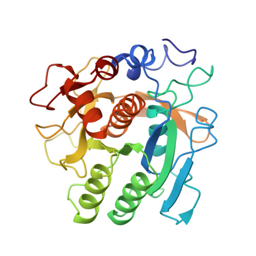
Crystal structure of thermitase at 1.4 A resolution.
Teplyakov, A.V., Kuranova, I.P., Harutyunyan, E.H., Vainshtein, B.K., Frommel, C., Hohne, W.E., Wilson, K.S.(1990) J Mol Biol 214: 261-279
- PubMed: 2196375
- DOI: https://doi.org/10.1016/0022-2836(90)90160-n
- Primary Citation of Related Structures:
1THM - PubMed Abstract:
The crystal structure of thermitase, a subtilisin-type serine proteinase from Thermoactinomyces vulgaris, was determined by X-ray diffraction at 1.4 A resolution. The structure was solved by a combination of molecular and isomorphous replacement. The starting model was that of subtilisin BPN' from the Protein Data Bank, determined at 2.5 A resolution. The high-resolution refinement was based on data collected using synchrotron radiation with a Fuji image plate as detector. The model of thermitase refined to a conventional R factor of 14.9% and contains 1997 protein atoms, 182 water molecules and two Ca ions. The tertiary structure of thermitase is similar to that of the other subtilisins although there are some significant differences in detail. Comparison with subtilisin BPN' revealed two major structural differences. The N-terminal region in thermitase, which is absent in subtilisin BPN', forms a number of contacts with the tight Ca2+ binding site and indeed provides the very tight binding of the Ca ion. In thermitase the loop of residues 60 to 65 forms an additional (10) beta-strand of the central beta-sheet and the second Ca2+ binding site that has no equivalent in the subtilisin BPN' structure. The observed differences in the Ca2+ binding and the increased number of ionic and aromatic interactions in thermitase are likely sources of the enhanced stability of thermitase.
Organizational Affiliation:
Institute of Crystallography, Academy of Sciences of the U.S.S.R., Moscow.