1TFU

phosphopantetheine adenylyltransferase from Mycobacterium tuberculosis


Experimental Data Snapshot

  • Method: X-RAY DIFFRACTION
  • Resolution: 1.99 Å
  • R-Value Free: 0.258 
  • R-Value Work: 0.229 
  • R-Value Observed: 0.229 

wwPDB Validation   3D Report Full Report


This is version 1.4 of the entry. See complete history


Literature

Substrate-induced asymmetry and channel closure revealed by the apoenzyme structure of Mycobacterium tuberculosis phosphopantetheine adenylyltransferase.

Morris, V.K.Izard, T.

(2004) Protein Sci 13: 2547-2552

  • DOI: https://doi.org/10.1110/ps.04816904
  • Primary Citation of Related Structures:  
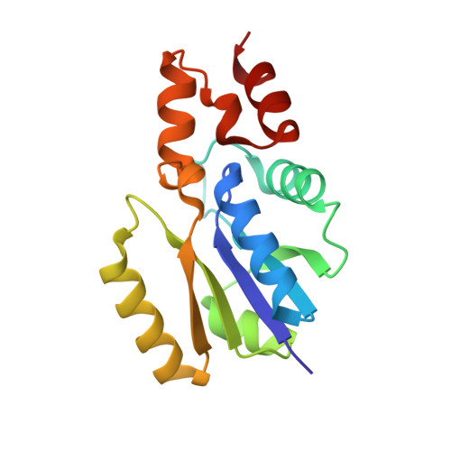
    1TFU

  • PubMed Abstract: 

    Phosphopantetheine adenylyltransferase (PPAT) catalyzes the penultimate step in prokaryotic coenzyme A (CoA) biosynthesis, directing the transfer of an adenylyl group from ATP to 4'-phosphopantetheine (Ppant) to yield dephospho-CoA (dPCoA). The crystal structures of Escherichia coli PPAT bound to its substrates, product, and inhibitor revealed an allosteric hexameric enzyme with half-of-sites reactivity, and established an in-line displacement catalytic mechanism. To provide insight into the mechanism of ligand binding we solved the apoenzyme (Apo) crystal structure of PPAT from Mycobacterium tuberculosis. In its Apo form, PPAT is a symmetric hexamer with an open solvent channel. However, ligand binding provokes asymmetry and alters the structure of the solvent channel, so that ligand binding becomes restricted to one trimer.


  • Organizational Affiliation

    Department of Hematology-Oncology, St. Jude Children's Research Hospital, Memphis TN 38105, USA.


Macromolecules
Find similar proteins by:  (by identity cutoff)  |  3D Structure
Entity ID: 1
MoleculeChains Sequence LengthOrganismDetailsImage
Phosphopantetheine adenylyltransferase157Mycobacterium tuberculosisMutation(s): 0 
Gene Names: COADKDTBRV2965CMT3043MTCY349.22U0002EMB2989C
EC: 2.7.7.3
UniProt
Find proteins for P9WPA5 (Mycobacterium tuberculosis (strain ATCC 25618 / H37Rv))
Explore P9WPA5 
Go to UniProtKB:  P9WPA5
Entity Groups  
Sequence Clusters30% Identity50% Identity70% Identity90% Identity95% Identity100% Identity
UniProt GroupP9WPA5
Sequence Annotations
Expand
  • Reference Sequence
Experimental Data & Validation

Experimental Data

  • Method: X-RAY DIFFRACTION
  • Resolution: 1.99 Å
  • R-Value Free: 0.258 
  • R-Value Work: 0.229 
  • R-Value Observed: 0.229 
  • Space Group: H 3 2
Unit Cell:
Length ( Å )Angle ( ˚ )
a = 98.623α = 90
b = 98.623β = 90
c = 114.811γ = 120
Software Package:
Software NamePurpose
CNSrefinement

Structure Validation

View Full Validation Report


Entry History 

Deposition Data

Revision History  (Full details and data files)

  • Version 1.0: 2004-09-14
    Type: Initial release
  • Version 1.1: 2008-04-30
    Changes: Version format compliance
  • Version 1.2: 2011-07-13
    Changes: Derived calculations, Version format compliance
  • Version 1.3: 2011-11-16
    Changes: Atomic model
  • Version 1.4: 2024-02-14
    Changes: Data collection, Database references