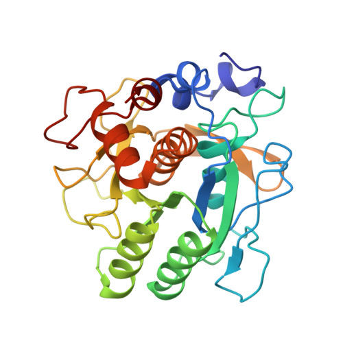
1TEC

CRYSTALLOGRAPHIC REFINEMENT BY INCORPORATION OF MOLECULAR DYNAMICS. THE THERMOSTABLE SERINE PROTEASE THERMITASE COMPLEXED WITH EGLIN-C


Experimental Data Snapshot

  • Method: X-RAY DIFFRACTION
  • Resolution: 2.20 Å

wwPDB Validation   3D Report Full Report


This is version 1.3 of the entry. See complete history


Literature

Crystallographic refinement by incorporation of molecular dynamics: thermostable serine protease thermitase complexed with eglin c.

Gros, P.Fujinaga, M.Dijkstra, B.W.Kalk, K.H.Hol, W.G.

(1989) Acta Crystallogr B 45: 488-499

  • DOI: https://doi.org/10.1107/s0108768189006038
  • Primary Citation of Related Structures:  
    1TEC

  • PubMed Abstract: 

    In order to investigate the principles of protein thermostability, the crystal structure of thermitase from Thermoactinomyces vulgaris, a thermostable member of the subtilisin family of serine proteases, has been determined in a complex with eglin c. Eglin c is a serine protease inhibitor from the leech Hirudo medicinalis. After data collection with a television area-detector diffractometer and initial structure solution by molecular-replacement methods, crystallographic refinement proceeded with incorporation of molecular-dynamics techniques. It appeared that this refinement procedure has a large convergence radius with movements of more than 5 A for many atoms. Two procedures for the crystallographic molecular-dynamics refinement have been tested. They differed mainly in time span and weight on the X-ray 'energy'. The best results were obtained with a procedure which allowed the molecular-dynamics technique to search a large area in conformational space by having less weight on the X-ray restraints and allowing more time. The use of molecular-dynamics refinement considerably simplified the laborious and difficult task of fitting the model in its electron density during the refinement process. The final crystallographic R factor is 17.9% at 2.2 A resolution.


  • Organizational Affiliation

    Department of Chemistry, University of Groningen, The Netherlands.


Macromolecules
Find similar proteins by:  (by identity cutoff)  |  3D Structure
Entity ID: 1
MoleculeChains Sequence LengthOrganismDetailsImage
THERMITASEA [auth E]279Thermoactinomyces vulgarisMutation(s): 0 
EC: 3.4.21.66
UniProt
Find proteins for P04072 (Thermoactinomyces vulgaris)
Explore P04072 
Go to UniProtKB:  P04072
Entity Groups  
Sequence Clusters30% Identity50% Identity70% Identity90% Identity95% Identity100% Identity
UniProt GroupP04072
Sequence Annotations
Expand
  • Reference Sequence
Find similar proteins by:  (by identity cutoff)  |  3D Structure
Entity ID: 2
MoleculeChains Sequence LengthOrganismDetailsImage
EGLIN CB [auth I]70Hirudo medicinalisMutation(s): 0 
UniProt
Find proteins for P01051 (Hirudo medicinalis)
Explore P01051 
Go to UniProtKB:  P01051
Entity Groups  
Sequence Clusters30% Identity50% Identity70% Identity90% Identity95% Identity100% Identity
UniProt GroupP01051
Sequence Annotations
Expand
  • Reference Sequence
Experimental Data & Validation

Experimental Data

  • Method: X-RAY DIFFRACTION
  • Resolution: 2.20 Å
  • Space Group: P 21 21 21
Unit Cell:
Length ( Å )Angle ( ˚ )
a = 63.25α = 90
b = 72.1β = 90
c = 89.25γ = 90
Software Package:
Software NamePurpose
GROMOSrefinement

Structure Validation

View Full Validation Report


Entry History 

Deposition Data

Revision History  (Full details and data files)

  • Version 1.0: 1989-10-15
    Type: Initial release
  • Version 1.1: 2008-03-24
    Changes: Version format compliance
  • Version 1.2: 2011-07-13
    Changes: Version format compliance
  • Version 1.3: 2024-02-14
    Changes: Data collection, Database references, Derived calculations, Other