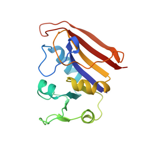
Expression, characterization and crystallographic analysis of telluromethionyl dihydrofolate reductase.
Boles, J.O., Lewinski, K., Kunckle, M.G., Hatada, M., Lebioda, L., Dunlap, R.B., Odom, J.D.(1995) Acta Crystallogr D Biol Crystallogr 51: 731-739
- PubMed: 15299803
- DOI: https://doi.org/10.1107/S0907444995001156
- Primary Citation of Related Structures:
1TDR - PubMed Abstract:
Selenomethionine-containing proteins analyzed by multi-wavelength anomalous diffraction provide a facile means of addressing the phase problem, whose solution is necessary to determine protein structures by X-ray crystallography [Hendrickson (1991). Science, 254, 51-58]. Since this method requires synchrotron radiation, we sought to incorporate a true heavy atom into protein, allowing the solution of the phase problem by more traditional methods of data collection. Media containing TeMet alone or TeMet with low levels of Met failed to sustain growth of a methione auxotroph of Escherichia coli carrying the dihydrofolate reductase expression vector. Growth of the organism to stationary phase and incorporation of TeMet was observed when the culture was initiated in media containing minimal Met levels and TeMet was added after induction with isopropyl-1-thio-beta-D-galactopyranoside. The purified enzyme exhibited properties similar to those of the native enzyme. Atomic absorption spectroscopy and amino-acid analysis indicated that 40% of the methionines were replaced with TeMet. Sequence analysis did not indicate significant levels of replacement in the first three sites (1, 16 and 20), suggesting that TeMet was present only in the last two sites (42 and 92). Crystals of this enzyme were grown in the presence of methotrexate and were isomorphous with crystals of wild-type dihydrofolate reductase. Difference Fourier maps and restrained least-squares refinement showed no substitution at the first three methionines, while incorporation was seen at positions 42 and 92.
Organizational Affiliation:
Department of Chemistry, Tennessee Technological University, Cookeville, TN 38505, USA.