1TDQ

Structural basis for the interactions between tenascins and the C-type lectin domains from lecticans: evidence for a cross-linking role for tenascins


Experimental Data Snapshot

  • Method: X-RAY DIFFRACTION
  • Resolution: 2.60 Å
  • R-Value Free: 0.234 
  • R-Value Work: 0.209 
  • R-Value Observed: 0.210 

wwPDB Validation   3D Report Full Report


This is version 1.4 of the entry. See complete history


Literature

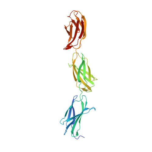
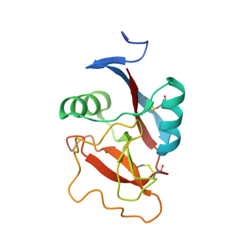
Structural basis for interactions between tenascins and lectican C-type lectin domains: evidence for a crosslinking role for tenascins

Lundell, A.Olin, A.I.Moergelin, M.al-Karadaghi, S.Aspberg, A.Logan, D.T.

(2004) Structure 12: 1495-1506

  • DOI: https://doi.org/10.1016/j.str.2004.05.021
  • Primary Citation of Related Structures:  
    1TDQ

  • PubMed Abstract: 

    The C-terminal G3 domains of lecticans mediate crosslinking to diverse extracellular matrix (ECM) proteins during ECM assembly, through their C-type lectin (CLD) subdomains. The structure of the rat aggrecan CLD in a Ca(2+)-dependent complex with fibronectin type III repeats 3-5 of rat tenascin-R provides detailed support for such crosslinking. The CLD loops bind Ca2+ like other CLDs, but no carbohydrate binding is observed or possible. This is thus the first example of a direct Ca(2+)-dependent protein-protein interaction of a CLD. Surprisingly, tenascin-R does not coordinate the Ca2+ ions directly. Electron microscopy confirms that full-length tenascin-R and tenascin-C crosslink hyaluronan-aggrecan complexes. The results are significant for the binding of all lectican CLDs to tenascin-R and tenascin-C. Comparison of the protein interaction surface with that of P-selectin in complex with the PGSL-1 peptide suggests that direct protein-protein interactions of Ca(2+)-binding CLDs may be more widespread than previously appreciated.


  • Organizational Affiliation

    Department of Molecular Biophysics, Lund University, Box 124, S-221 00 Lund, Sweden.


Macromolecules
Find similar proteins by:  (by identity cutoff)  |  3D Structure
Entity ID: 1
MoleculeChains Sequence LengthOrganismDetailsImage
Tenascin-R283Rattus norvegicusMutation(s): 0 
UniProt
Find proteins for Q05546 (Rattus norvegicus)
Explore Q05546 
Go to UniProtKB:  Q05546
Entity Groups  
Sequence Clusters30% Identity50% Identity70% Identity90% Identity95% Identity100% Identity
UniProt GroupQ05546
Sequence Annotations
Expand
  • Reference Sequence
Find similar proteins by:  (by identity cutoff)  |  3D Structure
Entity ID: 2
MoleculeChains Sequence LengthOrganismDetailsImage
Aggrecan core protein130Rattus norvegicusMutation(s): 0 
Gene Names: AGC1AGC
UniProt
Find proteins for P07897 (Rattus norvegicus)
Explore P07897 
Go to UniProtKB:  P07897
Entity Groups  
Sequence Clusters30% Identity50% Identity70% Identity90% Identity95% Identity100% Identity
UniProt GroupP07897
Sequence Annotations
Expand
  • Reference Sequence
Experimental Data & Validation

Experimental Data

  • Method: X-RAY DIFFRACTION
  • Resolution: 2.60 Å
  • R-Value Free: 0.234 
  • R-Value Work: 0.209 
  • R-Value Observed: 0.210 
  • Space Group: P 21 21 2
Unit Cell:
Length ( Å )Angle ( ˚ )
a = 112.52α = 90
b = 86.42β = 90
c = 57.6γ = 90
Software Package:
Software NamePurpose
REFMACrefinement
MOSFLMdata reduction
CCP4data scaling
SHARPphasing

Structure Validation

View Full Validation Report


Entry History 

Deposition Data

Revision History  (Full details and data files)

  • Version 1.0: 2004-08-31
    Type: Initial release
  • Version 1.1: 2008-04-30
    Changes: Version format compliance
  • Version 1.2: 2011-07-13
    Changes: Advisory, Version format compliance
  • Version 1.3: 2018-03-07
    Changes: Data collection
  • Version 1.4: 2021-03-31
    Changes: Database references, Derived calculations, Source and taxonomy