1TC2

TERNARY SUBSTRATE COMPLEX OF THE HYPOXANTHINE PHOSPHORIBOSYLTRANSFERASE FROM TRYPANOSOMA CRUZI


Experimental Data Snapshot

  • Method: X-RAY DIFFRACTION
  • Resolution: 1.81 Å
  • R-Value Free: 0.232 
  • R-Value Work: 0.183 
  • R-Value Observed: 0.191 

wwPDB Validation   3D Report Full Report


Ligand Structure Quality Assessment 


This is version 1.6 of the entry. See complete history


Literature

Approaching the transition state in the crystal structure of a phosphoribosyltransferase.

Focia, P.J.Craig III, S.P.Eakin, A.E.

(1998) Biochemistry 37: 17120-17127

  • DOI: https://doi.org/10.1021/bi9821465
  • Primary Citation of Related Structures:  
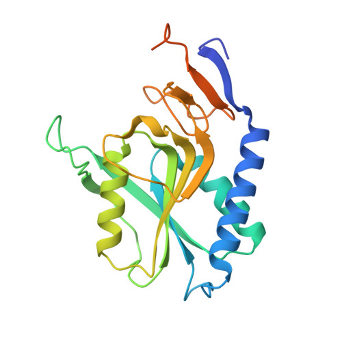
    1TC2

  • PubMed Abstract: 

    Hypoxanthine phosphoribosyltransferase (HPRT) salvages 6-oxopurine bases in the nucleotide metabolic pathway. The 1.8 A crystal structure of an asymmetric dimer of the HPRT from the protozoan parasite Trypanosoma cruzi was determined in a ternary complex with the primary substrate phosphoribosylpyrophosphate (PRPP) and an analogue of the substrate hypoxanthine, revealing both open and closed active site conformations. The ligands are positioned for in-line nucleophilic attack at the PRPP ribose C1' by two metal ions which straddle the pyrophosphate leaving group. The structure provides the first evidence for the involvement of two metal ions in the HPRT-catalyzed reaction, and structural details further suggest the mechanism may proceed via SN2-type chemistry. The closed conformation reveals the structural roles for invariant flexible loop residues Ser103 and Tyr104 and supports a role for the loop in the liberation of pyrophosphate. The pre-transition state structure is valuable for understanding the enzyme mechanism, as well as providing a foundation for antiparasite drug design efforts against T. cruzi, which causes Chagas' disease in humans. Additionally, the structure illuminates the molecular basis of three inherited mutations in the human HPRT leading to Lesch-Nyhan syndrome (D193N) or gout (S103R or S109L), as the homologous residues in the trypanosomal enzyme contribute to the previously unrecognized Mg2+ ion binding site and to the formation of the closed flexible loop, respectively.


  • Organizational Affiliation

    Laboratory of Molecular Parasitology & Drug Design, School of Pharmacy, University of North Carolina, Chapel Hill 27599-7360, USA.


Macromolecules
Find similar proteins by:  (by identity cutoff)  |  3D Structure
Entity ID: 1
MoleculeChains Sequence LengthOrganismDetailsImage
PROTEIN (HYPOXANTHINE PHOSPHORIBOSYLTRANSFERASE)
A, B
221Trypanosoma cruziMutation(s): 3 
EC: 2.4.2.8
UniProt
Find proteins for Q4DRC4 (Trypanosoma cruzi (strain CL Brener))
Explore Q4DRC4 
Go to UniProtKB:  Q4DRC4
Entity Groups  
Sequence Clusters30% Identity50% Identity70% Identity90% Identity95% Identity100% Identity
UniProt GroupQ4DRC4
Sequence Annotations
Expand
  • Reference Sequence
Small Molecules
Ligands 4 Unique
IDChains Name / Formula / InChI Key2D Diagram3D Interactions
PRP
Query on PRP

Download Ideal Coordinates CCD File 
F [auth A],
J [auth B]
1-O-pyrophosphono-5-O-phosphono-alpha-D-ribofuranose
C5 H13 O14 P3
PQGCEDQWHSBAJP-TXICZTDVSA-N
7HP
Query on 7HP

Download Ideal Coordinates CCD File 
E [auth A],
I [auth B]
7-HYDROXY-PYRAZOLO[4,3-D]PYRIMIDINE
C5 H4 N4 O
JFZSDNLQDTYVEE-UHFFFAOYSA-N
MN
Query on MN

Download Ideal Coordinates CCD File 
H [auth B]MANGANESE (II) ION
Mn
WAEMQWOKJMHJLA-UHFFFAOYSA-N
MG
Query on MG

Download Ideal Coordinates CCD File 
C [auth A],
D [auth A],
G [auth B]
MAGNESIUM ION
Mg
JLVVSXFLKOJNIY-UHFFFAOYSA-N
Experimental Data & Validation

Experimental Data

  • Method: X-RAY DIFFRACTION
  • Resolution: 1.81 Å
  • R-Value Free: 0.232 
  • R-Value Work: 0.183 
  • R-Value Observed: 0.191 
  • Space Group: P 1 21 1
Unit Cell:
Length ( Å )Angle ( ˚ )
a = 39.42α = 90
b = 101.76β = 94.23
c = 51.92γ = 90
Software Package:
Software NamePurpose
AMoREphasing
X-PLORrefinement
DENZOdata reduction
SCALEPACKdata scaling

Structure Validation

View Full Validation Report


Ligand Structure Quality Assessment 


Entry History 

Deposition Data

Revision History  (Full details and data files)

  • Version 1.0: 2000-03-08
    Type: Initial release
  • Version 1.1: 2008-04-27
    Changes: Version format compliance
  • Version 1.2: 2011-07-13
    Changes: Version format compliance
  • Version 1.3: 2018-01-24
    Changes: Database references
  • Version 1.4: 2018-01-31
    Changes: Database references
  • Version 1.5: 2020-07-29
    Type: Remediation
    Reason: Carbohydrate remediation
    Changes: Data collection, Database references, Derived calculations, Structure summary
  • Version 1.6: 2023-08-23
    Changes: Data collection, Database references, Refinement description, Structure summary