1T9G

Structure of the human MCAD:ETF complex


Experimental Data Snapshot

  • Method: X-RAY DIFFRACTION
  • Resolution: 2.90 Å
  • R-Value Free: 0.263 
  • R-Value Work: 0.191 
  • R-Value Observed: 0.195 

wwPDB Validation   3D Report Full Report


Ligand Structure Quality Assessment 


This is version 1.3 of the entry. See complete history


Literature

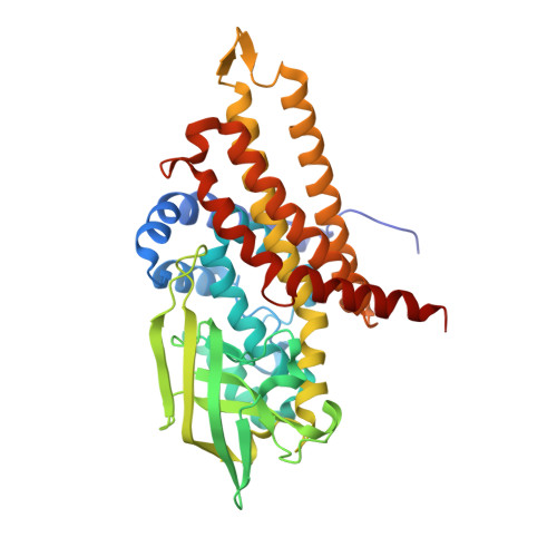
Extensive domain motion and electron transfer in the human electron transferring flavoprotein-medium chain Acyl-CoA dehydrogenase complex

Toogood, H.S.van Thiel, A.Basran, J.Sutcliffe, M.J.Scrutton, N.S.Leys, D.

(2004) J Biol Chem 279: 32904-32912

  • DOI: https://doi.org/10.1074/jbc.M404884200
  • Primary Citation of Related Structures:  
    1T9G

  • PubMed Abstract: 

    The crystal structure of the human electron transferring flavoprotein (ETF).medium chain acyl-CoA dehydrogenase (MCAD) complex reveals a dual mode of protein-protein interaction, imparting both specificity and promiscuity in the interaction of ETF with a range of structurally distinct primary dehydrogenases. ETF partitions the functions of partner binding and electron transfer between (i) the recognition loop, which acts as a static anchor at the ETF.MCAD interface, and (ii) the highly mobile redox active FAD domain. Together, these enable the FAD domain of ETF to sample a range of conformations, some compatible with fast interprotein electron transfer. Disorders in amino acid or fatty acid catabolism can be attributed to mutations at the protein-protein interface. Crucially, complex formation triggers mobility of the FAD domain, an induced disorder that contrasts with general models of protein-protein interaction by induced fit mechanisms. The subsequent interfacial motion in the MCAD.ETF complex is the basis for the interaction of ETF with structurally diverse protein partners. Solution studies using ETF and MCAD with mutations at the protein-protein interface support this dynamic model and indicate ionic interactions between MCAD Glu(212) and ETF Arg alpha(249) are likely to transiently stabilize productive conformations of the FAD domain leading to enhanced electron transfer rates between both partners.


  • Organizational Affiliation

    Department of Biochemistry, University of Leicester, Leicester LE1 7RH, United Kingdom.


Macromolecules
Find similar proteins by:  (by identity cutoff)  |  3D Structure
Entity ID: 1
MoleculeChains Sequence LengthOrganismDetailsImage
Acyl-CoA dehydrogenase, medium-chain specific, mitochondrial
A, B, C, D
396Homo sapiensMutation(s): 0 
Gene Names: ACADM
EC: 1.3.99.3
UniProt & NIH Common Fund Data Resources
Find proteins for P11310 (Homo sapiens)
Explore P11310 
Go to UniProtKB:  P11310
PHAROS:  P11310
GTEx:  ENSG00000117054 
Entity Groups  
Sequence Clusters30% Identity50% Identity70% Identity90% Identity95% Identity100% Identity
UniProt GroupP11310
Sequence Annotations
Expand
  • Reference Sequence
Find similar proteins by:  (by identity cutoff)  |  3D Structure
Entity ID: 2
MoleculeChains Sequence LengthOrganismDetailsImage
Electron transfer flavoprotein alpha-subunit, mitochondrialE [auth R]333Homo sapiensMutation(s): 0 
Gene Names: ETFA
UniProt & NIH Common Fund Data Resources
Find proteins for P13804 (Homo sapiens)
Explore P13804 
Go to UniProtKB:  P13804
PHAROS:  P13804
GTEx:  ENSG00000140374 
Entity Groups  
Sequence Clusters30% Identity50% Identity70% Identity90% Identity95% Identity100% Identity
UniProt GroupP13804
Sequence Annotations
Expand
  • Reference Sequence
Find similar proteins by:  (by identity cutoff)  |  3D Structure
Entity ID: 3
MoleculeChains Sequence LengthOrganismDetailsImage
Electron transfer flavoprotein beta-subunitF [auth S]255Homo sapiensMutation(s): 0 
Gene Names: ETFB
UniProt & NIH Common Fund Data Resources
Find proteins for P38117 (Homo sapiens)
Explore P38117 
Go to UniProtKB:  P38117
PHAROS:  P38117
GTEx:  ENSG00000105379 
Entity Groups  
Sequence Clusters30% Identity50% Identity70% Identity90% Identity95% Identity100% Identity
UniProt GroupP38117
Sequence Annotations
Expand
  • Reference Sequence
Experimental Data & Validation

Experimental Data

  • Method: X-RAY DIFFRACTION
  • Resolution: 2.90 Å
  • R-Value Free: 0.263 
  • R-Value Work: 0.191 
  • R-Value Observed: 0.195 
  • Space Group: P 21 21 21
Unit Cell:
Length ( Å )Angle ( ˚ )
a = 94.333α = 90
b = 101.322β = 90
c = 244.785γ = 90
Software Package:
Software NamePurpose
REFMACrefinement
DENZOdata reduction
SCALEPACKdata scaling
AMoREphasing

Structure Validation

View Full Validation Report


Ligand Structure Quality Assessment 


Entry History 

Deposition Data

Revision History  (Full details and data files)

  • Version 1.0: 2004-06-08
    Type: Initial release
  • Version 1.1: 2008-04-30
    Changes: Version format compliance
  • Version 1.2: 2011-07-13
    Changes: Version format compliance
  • Version 1.3: 2024-02-14
    Changes: Data collection, Database references, Derived calculations