1T2V

Structural basis of phospho-peptide recognition by the BRCT domain of BRCA1, structure with phosphopeptide


Experimental Data Snapshot

  • Method: X-RAY DIFFRACTION
  • Resolution: 3.30 Å
  • R-Value Free: 0.301 
  • R-Value Work: 0.258 
  • R-Value Observed: 0.261 

wwPDB Validation   3D Report Full Report


This is version 1.2 of the entry. See complete history


Literature

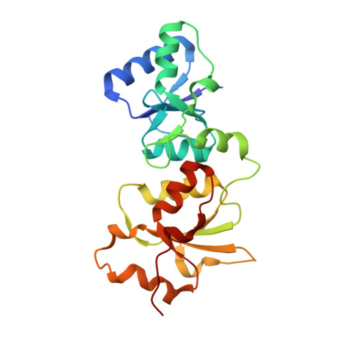
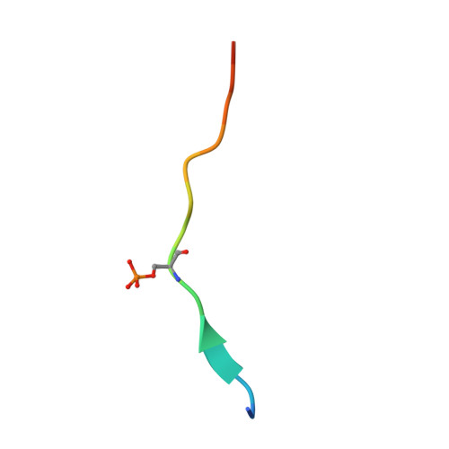
Structural basis of phosphopeptide recognition by the BRCT domain of BRCA1

Williams, R.S.Lee, M.S.Hau, D.D.Glover, J.N.M.

(2004) Nat Struct Mol Biol 11: 519-525

  • DOI: https://doi.org/10.1038/nsmb776
  • Primary Citation of Related Structures:  
    1T2U, 1T2V

  • PubMed Abstract: 

    The BRCT repeats in BRCA1 are essential for its tumor suppressor activity and interact with phosphorylated protein targets containing the sequence pSer-X-X-Phe, where X indicates any residue. The structure of the tandem BRCA1 BRCT repeats bound to an optimized phosphopeptide reveals that the N-terminal repeat harbors a conserved BRCT phosphoserine-binding pocket, while the interface between the repeats forms a hydrophobic groove that recognizes the phenylalanine. Crystallographic and biochemical data suggest that the structural integrity of both binding sites is essential for peptide recognition. The diminished peptide-binding capacity observed for cancer-associated BRCA1-BRCT variants may explain the enhanced cancer risks associated with these mutations.


  • Organizational Affiliation

    Department of Biochemistry, University of Alberta, Edmonton, Alberta, T6G 2H7, Canada.


Macromolecules
Find similar proteins by:  (by identity cutoff)  |  3D Structure
Entity ID: 1
MoleculeChains Sequence LengthOrganismDetailsImage
Breast cancer type 1 susceptibility protein
A, B, C, D, E
214Homo sapiensMutation(s): 0 
Gene Names: BRCA1
UniProt & NIH Common Fund Data Resources
Find proteins for P38398 (Homo sapiens)
Explore P38398 
Go to UniProtKB:  P38398
PHAROS:  P38398
GTEx:  ENSG00000012048 
Entity Groups  
Sequence Clusters30% Identity50% Identity70% Identity90% Identity95% Identity100% Identity
UniProt GroupP38398
Sequence Annotations
Expand
  • Reference Sequence

Find similar proteins by:  Sequence   |   3D Structure  

Entity ID: 2
MoleculeChains Sequence LengthOrganismDetailsImage
BRCTide-7PS
F, G, H, I, J
16N/AMutation(s): 0 
Entity Groups  
Sequence Clusters30% Identity50% Identity70% Identity90% Identity95% Identity100% Identity
Sequence Annotations
Expand
  • Reference Sequence
Small Molecules
Modified Residues  1 Unique
IDChains TypeFormula2D DiagramParent
SEP
Query on SEP
F, G, H, I, J
L-PEPTIDE LINKINGC3 H8 N O6 PSER
Experimental Data & Validation

Experimental Data

  • Method: X-RAY DIFFRACTION
  • Resolution: 3.30 Å
  • R-Value Free: 0.301 
  • R-Value Work: 0.258 
  • R-Value Observed: 0.261 
  • Space Group: C 2 2 21
Unit Cell:
Length ( Å )Angle ( ˚ )
a = 97.117α = 90
b = 138.357β = 90
c = 198.266γ = 90
Software Package:
Software NamePurpose
REFMACrefinement
DENZOdata reduction
SCALEPACKdata scaling
MOLREPphasing

Structure Validation

View Full Validation Report


Entry History 

Deposition Data

Revision History  (Full details and data files)

  • Version 1.0: 2004-05-11
    Type: Initial release
  • Version 1.1: 2008-04-30
    Changes: Version format compliance
  • Version 1.2: 2011-07-13
    Changes: Advisory, Version format compliance