1SK4

crystal structure of the C-terminal peptidoglycan-binding domain of human peptidoglycan recognition protein Ialpha


Experimental Data Snapshot

  • Method: X-RAY DIFFRACTION
  • Resolution: 1.65 Å
  • R-Value Free: 0.212 
  • R-Value Work: 0.196 
  • R-Value Observed: 0.196 

wwPDB Validation   3D Report Full Report


This is version 1.4 of the entry. See complete history


Literature

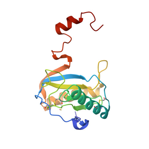
Crystal structure of the C-terminal peptidoglycan-binding domain of human peptidoglycan recognition protein Ialpha

Guan, R.Malchiodi, E.L.Qian, W.Schuck, P.Mariuzza, R.A.

(2004) J Biol Chem 279: 31873-31882

  • DOI: https://doi.org/10.1074/jbc.M404920200
  • Primary Citation of Related Structures:  
    1SK3, 1SK4

  • PubMed Abstract: 

    Peptidoglycan recognition proteins (PGRPs) are pattern recognition receptors of the innate immune system that bind, and in some cases hydrolyze, peptidoglycans (PGNs) on bacterial cell walls. These molecules, which are highly conserved from insects to mammals, participate in host defense against both Gram-positive and Gram-negative bacteria. We report the crystal structure of the C-terminal PGN-binding domain of human PGRP-Ialpha in two oligomeric states, monomer and dimer, to resolutions of 2.80 and 1.65 A, respectively. In contrast to PGRPs with PGN-lytic amidase activity, no zinc ion is present in the PGN-binding site of human PGRP-Ialpha. The structure reveals that PGRPs exhibit extensive topological variability in a large hydrophobic groove, located opposite the PGN-binding site, which may recognize host effector proteins or microbial ligands other than PGN. We also show that full-length PGRP-Ialpha comprises two tandem PGN-binding domains. These domains differ at most potential PGN-contacting positions, implying different fine specificities. Dimerization of PGRP-Ialpha, which occurs through three-dimensional domain swapping, is mediated by specific binding of sodium ions to a flexible hinge loop, stabilizing the conformation found in the dimer. We further demonstrate sodium-dependent dimerization of PGRP-Ialpha in solution, suggesting a possible mechanism for modulating PGRP activity through the formation of multivalent adducts.


  • Organizational Affiliation

    Center for Advanced Research in Biotechnology, W. M. Keck Laboratory for Structural Biology, University of Maryland Biotechnology Institute, Rockville, Maryland 20850, USA.


Macromolecules
Find similar proteins by:  (by identity cutoff)  |  3D Structure
Entity ID: 1
MoleculeChains Sequence LengthOrganismDetailsImage
Peptidoglycan recognition protein I-alpha163Homo sapiensMutation(s): 1 
Gene Names: PGRPIA
UniProt & NIH Common Fund Data Resources
Find proteins for Q96LB9 (Homo sapiens)
Explore Q96LB9 
Go to UniProtKB:  Q96LB9
PHAROS:  Q96LB9
GTEx:  ENSG00000159527 
Entity Groups  
Sequence Clusters30% Identity50% Identity70% Identity90% Identity95% Identity100% Identity
UniProt GroupQ96LB9
Sequence Annotations
Expand
  • Reference Sequence
Small Molecules
Ligands 1 Unique
IDChains Name / Formula / InChI Key2D Diagram3D Interactions
NA
Query on NA

Download Ideal Coordinates CCD File 
B [auth A]SODIUM ION
Na
FKNQFGJONOIPTF-UHFFFAOYSA-N
Modified Residues  1 Unique
IDChains TypeFormula2D DiagramParent
CSO
Query on CSO
A
L-PEPTIDE LINKINGC3 H7 N O3 SCYS
Experimental Data & Validation

Experimental Data

  • Method: X-RAY DIFFRACTION
  • Resolution: 1.65 Å
  • R-Value Free: 0.212 
  • R-Value Work: 0.196 
  • R-Value Observed: 0.196 
  • Space Group: P 32 2 1
Unit Cell:
Length ( Å )Angle ( ˚ )
a = 64.464α = 90
b = 64.464β = 90
c = 63.996γ = 120
Software Package:
Software NamePurpose
CNSrefinement
HKL-2000data reduction
SCALEPACKdata scaling
AMoREphasing

Structure Validation

View Full Validation Report


Entry History 

Deposition Data

Revision History  (Full details and data files)

  • Version 1.0: 2004-07-13
    Type: Initial release
  • Version 1.1: 2008-04-29
    Changes: Version format compliance
  • Version 1.2: 2011-07-13
    Changes: Derived calculations, Version format compliance
  • Version 1.3: 2023-08-23
    Changes: Data collection, Database references, Derived calculations, Refinement description
  • Version 1.4: 2023-11-15
    Changes: Data collection