1SGU

Comparing the Accumulation of Active Site and Non-active Site Mutations in the HIV-1 Protease


Experimental Data Snapshot

  • Method: X-RAY DIFFRACTION
  • Resolution: 1.90 Å
  • R-Value Free: 0.264 
  • R-Value Work: 0.220 
  • R-Value Observed: 0.220 

wwPDB Validation   3D Report Full Report


Ligand Structure Quality Assessment 


This is version 1.6 of the entry. See complete history


Literature

Comparing the Accumulation of Active- and Nonactive-Site Mutations in the HIV-1 Protease.

Clemente, J.C.Moose, R.E.Hemrajani, R.Whitford, L.R.Govindasamy, L.Reutzel, R.McKenna, R.Agbandje-McKenna, M.Goodenow, M.M.Dunn, B.M.

(2004) Biochemistry 43: 12141-12151

  • DOI: https://doi.org/10.1021/bi049459m
  • Primary Citation of Related Structures:  
    1SGU, 1SH9

  • PubMed Abstract: 

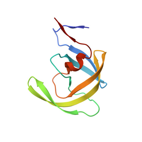
    Protease inhibitor resistance still poses one of the greatest challenges in treating HIV. To better design inhibitors able to target resistant proteases, a deeper understanding is needed of the effects of accumulating mutations and the contributions of active- and nonactive-site mutations to the resistance. We have engineered a series of variants containing the nonactive-site mutations M46I and I54V and the active-site mutation I84V. These mutations were added to a protease clone (V6) isolated from a pediatric patient on ritonavir therapy. This variant possessed the ritonavir-resistance-associated mutations in the active-site (V32I and V82A) and nonactive-site mutations (K20R, L33F, M36I, L63P, A71V, and L90M). The I84V mutation had the greatest effect on decreasing catalytic efficiency, 10-fold when compared to the pretherapy clone LAI. The decrease in catalytic efficiency was partially recovered by the addition of mutations M46I and I54V. The M46I and I54V were just as effective at decreasing inhibitor binding as the I84V mutation when compared to V6 and LAI. The V6(54/84) variant showed over 1000-fold decrease in inhibitor-binding strength to ritonavir, indinavir, and nelfinavir when compared to LAI and V6. Crystal-structure analysis of the V6(54/84) variant bound to ritonavir and indinavir shows structural changes in the 80's loops and active site, which lead to an enlarged binding cavity when compared to pretherapy structures in the Protein Data Bank. Structural changes are also seen in the 10's and 30's loops, which suggest possible changes in the dynamics of flap opening and closing.


  • Organizational Affiliation

    Department of Biochemistry and Molecular Biology, University of Florida College of Medicine, Gainesville, Florida 32610, USA. clemente@ufl.edu


Macromolecules
Find similar proteins by:  (by identity cutoff)  |  3D Structure
Entity ID: 1
MoleculeChains Sequence LengthOrganismDetailsImage
POL polyprotein
A, B
99Human immunodeficiency virus 1Mutation(s): 10 
EC: 3.4.23.16
UniProt
Find proteins for P03367 (Human immunodeficiency virus type 1 group M subtype B (isolate BRU/LAI))
Explore P03367 
Go to UniProtKB:  P03367
Entity Groups  
Sequence Clusters30% Identity50% Identity70% Identity90% Identity95% Identity100% Identity
UniProt GroupP03367
Sequence Annotations
Expand
  • Reference Sequence
Small Molecules
Ligands 1 Unique
IDChains Name / Formula / InChI Key2D Diagram3D Interactions
MK1
Query on MK1

Download Ideal Coordinates CCD File 
C [auth B]N-[2(R)-HYDROXY-1(S)-INDANYL]-5-[(2(S)-TERTIARY BUTYLAMINOCARBONYL)-4(3-PYRIDYLMETHYL)PIPERAZINO]-4(S)-HYDROXY-2(R)-PHENYLMETHYLPENTANAMIDE
C36 H47 N5 O4
CBVCZFGXHXORBI-PXQQMZJSSA-N
Binding Affinity Annotations 
IDSourceBinding Affinity
MK1 BindingDB:  1SGU Ki: min: 0.05, max: 398 (nM) from 35 assay(s)
Kd: 1.07 (nM) from 1 assay(s)
IC50: min: 0.36, max: 370 (nM) from 18 assay(s)
-TΔS: min: -7.40e+1, max: -2.80e+1 (kJ/mol) from 27 assay(s)
ΔH: min: -3.34e+1, max: 35.11 (kJ/mol) from 27 assay(s)
ΔG: min: -6.31e+1, max: -3.85e+1 (kJ/mol) from 28 assay(s)
PDBBind:  1SGU Ki: 4235 (nM) from 1 assay(s)
Binding MOAD:  1SGU Ki: 4235 (nM) from 1 assay(s)
Experimental Data & Validation

Experimental Data

  • Method: X-RAY DIFFRACTION
  • Resolution: 1.90 Å
  • R-Value Free: 0.264 
  • R-Value Work: 0.220 
  • R-Value Observed: 0.220 
  • Space Group: P 61
Unit Cell:
Length ( Å )Angle ( ˚ )
a = 61.573α = 90
b = 61.574β = 90
c = 84.109γ = 120
Software Package:
Software NamePurpose
CNSrefinement
SCALEPACKdata scaling
Omodel building

Structure Validation

View Full Validation Report


Ligand Structure Quality Assessment 


Entry History 

Deposition Data

Revision History  (Full details and data files)

  • Version 1.0: 2004-10-05
    Type: Initial release
  • Version 1.1: 2008-04-29
    Changes: Version format compliance
  • Version 1.2: 2011-07-13
    Changes: Version format compliance
  • Version 1.3: 2017-10-11
    Changes: Refinement description
  • Version 1.4: 2018-01-31
    Changes: Experimental preparation
  • Version 1.5: 2021-10-27
    Changes: Database references, Derived calculations
  • Version 1.6: 2024-02-14
    Changes: Data collection