1SBS

CRYSTAL STRUCTURE OF AN ANTI-HCG FAB


Experimental Data Snapshot

  • Method: X-RAY DIFFRACTION
  • Resolution: 2.00 Å
  • R-Value Free: 0.240 
  • R-Value Work: 0.180 
  • R-Value Observed: 0.180 

wwPDB Validation   3D Report Full Report


This is version 1.3 of the entry. See complete history


Literature

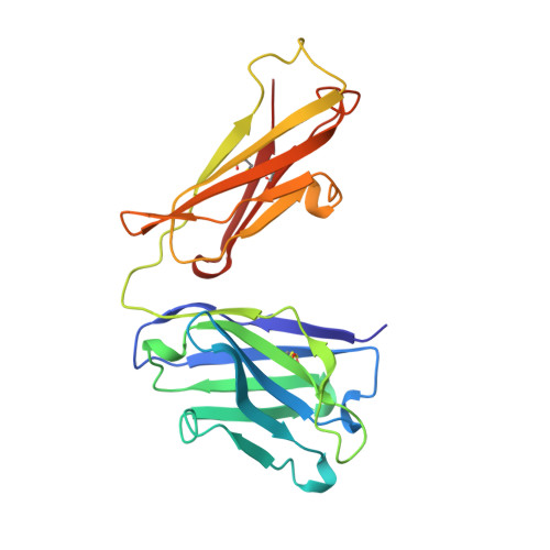
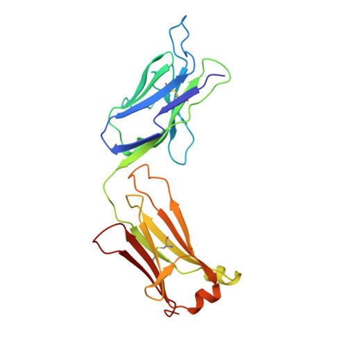
Structure of an Fab fragment against a C-terminal peptide of hCG at 2.0 A resolution.

Fotinou, C.Beauchamp, J.Emsley, P.deHaan, A.Schielen, W.J.Bos, E.Isaacs, N.W.

(1998) J Biol Chem 273: 22515-22518

  • DOI: https://doi.org/10.1074/jbc.273.35.22515
  • Primary Citation of Related Structures:  
    1SBS

  • PubMed Abstract: 

    3A2 is an antibody raised against human chorionic gonadotropin and recognizes a linear epitope on the C-terminal peptide of the human chorionic gonadotropin beta-subunit. Its three-dimensional structure has been determined to 2-A resolution using molecular replacement and refined to a conventional R-factor of 18.2%. The protein exhibits the typical immunoglobulin fold, and the model contains 944 ordered water molecules and one sulfate ion. A comparison of the complementarity-determining regions of the Fab3A2 with those from the Protein Data Bank following the canonical structure method reveals a canonical main chain conformation. This antibody belongs to the canonical structure class (combination of canonical conformations of the complementarity determining loops) that shows a preference for haptens and not for peptides. However, the shape of the surface of the antigen binding loops resembles that of an anti-peptide antibody.


  • Organizational Affiliation

    Department of Chemistry, University of Glasgow, Glasgow G12 8QQ, United Kingdom.


Macromolecules
Find similar proteins by:  (by identity cutoff)  |  3D Structure
Entity ID: 1
MoleculeChains Sequence LengthOrganismDetailsImage
MONOCLONAL ANTIBODY 3A2A [auth H]222Mus musculusMutation(s): 0 
Entity Groups  
Sequence Clusters30% Identity50% Identity70% Identity90% Identity95% Identity100% Identity
Sequence Annotations
Expand
  • Reference Sequence
Find similar proteins by:  (by identity cutoff)  |  3D Structure
Entity ID: 2
MoleculeChains Sequence LengthOrganismDetailsImage
MONOCLONAL ANTIBODY 3A2B [auth L]220Mus musculusMutation(s): 0 
Entity Groups  
Sequence Clusters30% Identity50% Identity70% Identity90% Identity95% Identity100% Identity
Sequence Annotations
Expand
  • Reference Sequence
Small Molecules
Ligands 1 Unique
IDChains Name / Formula / InChI Key2D Diagram3D Interactions
SO4
Query on SO4

Download Ideal Coordinates CCD File 
C [auth H]SULFATE ION
O4 S
QAOWNCQODCNURD-UHFFFAOYSA-L
Experimental Data & Validation

Experimental Data

  • Method: X-RAY DIFFRACTION
  • Resolution: 2.00 Å
  • R-Value Free: 0.240 
  • R-Value Work: 0.180 
  • R-Value Observed: 0.180 
  • Space Group: P 31 2 1
Unit Cell:
Length ( Å )Angle ( ˚ )
a = 74.8α = 90
b = 74.8β = 90
c = 198.205γ = 120
Software Package:
Software NamePurpose
DENZOdata reduction
ROTAVATA/AGROVATAdata reduction
X-PLORmodel building
X-PLORrefinement
CCP4data scaling
ROTAVATAdata scaling
X-PLORphasing

Structure Validation

View Full Validation Report


Entry History 

Deposition Data

Revision History  (Full details and data files)

  • Version 1.0: 1999-04-13
    Type: Initial release
  • Version 1.1: 2008-03-24
    Changes: Version format compliance
  • Version 1.2: 2011-07-13
    Changes: Derived calculations, Version format compliance
  • Version 1.3: 2023-08-09
    Changes: Database references, Derived calculations, Refinement description