1S5G

Structure of Scallop myosin S1 reveals a novel nucleotide conformation


Experimental Data Snapshot

  • Method: X-RAY DIFFRACTION
  • Resolution: 3.10 Å
  • R-Value Free: 0.269 
  • R-Value Work: 0.233 
  • R-Value Observed: 0.233 

wwPDB Validation   3D Report Full Report


Ligand Structure Quality Assessment 


This is version 1.3 of the entry. See complete history


Literature

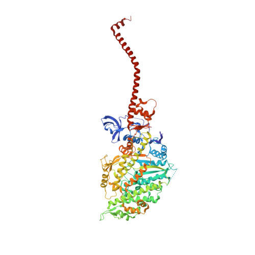
Myosin subfragment 1 structures reveal a partially bound nucleotide and a complex salt bridge that helps couple nucleotide and actin binding.

Risal, D.Gourinath, S.Himmel, D.M.Szent-Gyorgyi, A.G.Cohen, C.

(2004) Proc Natl Acad Sci U S A 101: 8930-8935

  • DOI: https://doi.org/10.1073/pnas.0403002101
  • Primary Citation of Related Structures:  
    1S5G, 1SR6

  • PubMed Abstract: 

    Structural studies of myosin have indicated some of the conformational changes that occur in this protein during the contractile cycle, and we have now observed a conformational change in a bound nucleotide as well. The 3.1-A x-ray structure of the scallop myosin head domain (subfragment 1) in the ADP-bound near-rigor state (lever arm =45 degrees to the helical actin axis) shows the diphosphate moiety positioned on the surface of the nucleotide-binding pocket, rather than deep within it as had been observed previously. This conformation strongly suggests a specific mode of entry and exit of the nucleotide from the nucleotide-binding pocket through the so-called "front door." In addition, using a variety of scallop structures, including a relatively high-resolution 2.75-A nucleotide-free near-rigor structure, we have identified a conserved complex salt bridge connecting the 50-kDa upper and N-terminal subdomains. This salt bridge is present only in crystal structures of muscle myosin isoforms that exhibit a strong reciprocal relationship (also known as coupling) between actin and nucleotide affinity.


  • Organizational Affiliation

    Rosenstiel Basic Medical Sciences Research Center, MS 029, Waltham, MA 02454-9110, USA.


Macromolecules
Find similar proteins by:  (by identity cutoff)  |  3D Structure
Entity ID: 1
MoleculeChains Sequence LengthOrganismDetailsImage
Myosin heavy chain, striated muscle840Argopecten irradiansMutation(s): 0 
UniProt
Find proteins for P24733 (Argopecten irradians)
Explore P24733 
Go to UniProtKB:  P24733
Entity Groups  
Sequence Clusters30% Identity50% Identity70% Identity90% Identity95% Identity100% Identity
UniProt GroupP24733
Sequence Annotations
Expand
  • Reference Sequence
Find similar proteins by:  (by identity cutoff)  |  3D Structure
Entity ID: 2
MoleculeChains Sequence LengthOrganismDetailsImage
Myosin regulatory light chain, striated adductor muscleB [auth Y]156Argopecten irradiansMutation(s): 0 
UniProt
Find proteins for P13543 (Argopecten irradians)
Explore P13543 
Go to UniProtKB:  P13543
Entity Groups  
Sequence Clusters30% Identity50% Identity70% Identity90% Identity95% Identity100% Identity
UniProt GroupP13543
Sequence Annotations
Expand
  • Reference Sequence
Find similar proteins by:  (by identity cutoff)  |  3D Structure
Entity ID: 3
MoleculeChains Sequence LengthOrganismDetailsImage
Myosin essential light chain, striated adductor muscleC [auth Z]156Argopecten irradiansMutation(s): 0 
UniProt
Find proteins for P07291 (Argopecten irradians)
Explore P07291 
Go to UniProtKB:  P07291
Entity Groups  
Sequence Clusters30% Identity50% Identity70% Identity90% Identity95% Identity100% Identity
UniProt GroupP07291
Sequence Annotations
Expand
  • Reference Sequence
Experimental Data & Validation

Experimental Data

  • Method: X-RAY DIFFRACTION
  • Resolution: 3.10 Å
  • R-Value Free: 0.269 
  • R-Value Work: 0.233 
  • R-Value Observed: 0.233 
  • Space Group: P 1 21 1
Unit Cell:
Length ( Å )Angle ( ˚ )
a = 84.051α = 90
b = 50.935β = 98.36
c = 161.59γ = 90
Software Package:
Software NamePurpose
DENZOdata reduction
SCALEPACKdata scaling
AMoREphasing
CNSrefinement

Structure Validation

View Full Validation Report


Ligand Structure Quality Assessment 


Entry History 

Deposition Data

Revision History  (Full details and data files)

  • Version 1.0: 2004-06-22
    Type: Initial release
  • Version 1.1: 2008-04-29
    Changes: Version format compliance
  • Version 1.2: 2011-07-13
    Changes: Version format compliance
  • Version 1.3: 2023-08-23
    Changes: Data collection, Database references, Derived calculations, Refinement description