1S0J

Trypanosoma cruzi trans-sialidase in complex with MuNANA (Michaelis complex)


Experimental Data Snapshot

  • Method: X-RAY DIFFRACTION
  • Resolution: 1.65 Å
  • R-Value Free: 0.194 
  • R-Value Work: 0.162 
  • R-Value Observed: 0.163 

wwPDB Validation   3D Report Full Report


Ligand Structure Quality Assessment 


This is version 1.4 of the entry. See complete history


Literature

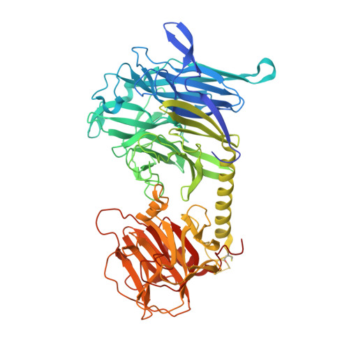
Structural Insights into the Catalytic Mechanism of Trypanosoma cruzi trans-Sialidase.

Amaya, M.F.Watts, A.G.Damager, I.Wehenkel, A.Nguyen, T.Buschiazzo, A.Paris, G.Frasch, A.C.Withers, S.G.Alzari, P.M.

(2004) Structure 12: 775-784

  • DOI: https://doi.org/10.1016/j.str.2004.02.036
  • Primary Citation of Related Structures:  
    1S0J

  • PubMed Abstract: 

    Sialidases are a superfamily of sialic-acid-releasing enzymes that are of significant interest due to their implication as virulence factors in the pathogenesis of a number of diseases. However, extensive studies of viral and microbial sialidases have failed to provide a comprehensive picture of their mechanistic properties, in part because the structures of competent enzyme-substrate complexes and reaction intermediates have never been described. Here we report these structures for the Trypanosoma cruzi trans-sialidase (TcTS), showing that catalysis by sialidases occurs via a similar mechanism to that of other retaining glycosidases, but with some intriguing differences that may have evolved in response to the substrate structure.


  • Organizational Affiliation

    Unité de Biochimie Structurale, CNRS URA 2185, Institut Pasteur, 25 rue du Dr. Roux, 75724 Paris, France.


Macromolecules
Find similar proteins by:  (by identity cutoff)  |  3D Structure
Entity ID: 1
MoleculeChains Sequence LengthOrganismDetailsImage
trans-sialidase648Trypanosoma cruziMutation(s): 8 
EC: 3.2.1.18
UniProt
Find proteins for Q26966 (Trypanosoma cruzi)
Explore Q26966 
Go to UniProtKB:  Q26966
Entity Groups  
Sequence Clusters30% Identity50% Identity70% Identity90% Identity95% Identity100% Identity
UniProt GroupQ26966
Sequence Annotations
Expand
  • Reference Sequence
Small Molecules
Ligands 1 Unique
IDChains Name / Formula / InChI Key2D Diagram3D Interactions
MUS
Query on MUS

Download Ideal Coordinates CCD File 
B [auth A]4-METHYL-2-OXO-2H-CHROMEN-7-YL 5-(ACETYLAMINO)-3,5-DIDEOXY-L-ERYTHRO-NON-2-ULOPYRANOSIDONIC ACID
C21 H25 N O11
KKDWIUJBUSOPGC-GKHMPSLRSA-N
Experimental Data & Validation

Experimental Data

  • Method: X-RAY DIFFRACTION
  • Resolution: 1.65 Å
  • R-Value Free: 0.194 
  • R-Value Work: 0.162 
  • R-Value Observed: 0.163 
  • Space Group: P 1 21 1
Unit Cell:
Length ( Å )Angle ( ˚ )
a = 54.373α = 90
b = 129.423β = 108.33
c = 54.234γ = 90
Software Package:
Software NamePurpose
REFMACrefinement
DENZOdata reduction
SCALEPACKdata scaling
AMoREphasing

Structure Validation

View Full Validation Report


Ligand Structure Quality Assessment 


Entry History 

Deposition Data

Revision History  (Full details and data files)

  • Version 1.0: 2004-05-18
    Type: Initial release
  • Version 1.1: 2008-04-29
    Changes: Version format compliance
  • Version 1.2: 2011-07-13
    Changes: Version format compliance
  • Version 1.3: 2021-10-27
    Changes: Database references, Derived calculations
  • Version 1.4: 2023-08-23
    Changes: Data collection, Refinement description