1RZ9

Crystal Structure of AAV Rep complexed with the Rep-binding sequence


Experimental Data Snapshot

  • Method: X-RAY DIFFRACTION
  • Resolution: 3.10 Å
  • R-Value Free: 0.315 
  • R-Value Work: 0.286 
  • R-Value Observed: 0.286 

wwPDB Validation   3D Report Full Report


This is version 1.3 of the entry. See complete history


Literature

The nuclease domain of adeno-associated virus rep coordinates replication initiation using two distinct DNA recognition interfaces.

Hickman, A.B.Ronning, D.R.Perez, Z.N.Kotin, R.M.Dyda, F.

(2004) Mol Cell 13: 403-414

  • DOI: https://doi.org/10.1016/s1097-2765(04)00023-1
  • Primary Citation of Related Structures:  
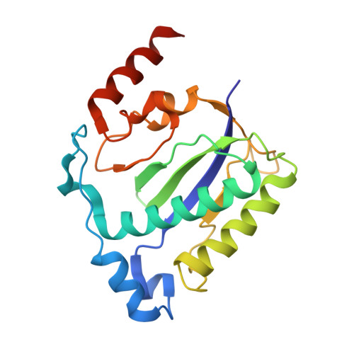
    1RZ9, 1UUT

  • PubMed Abstract: 

    Integration into a particular location in human chromosomes is a unique property of the adeno-associated virus (AAV). This reaction requires the viral Rep protein and AAV origin sequences. To understand how Rep recognizes DNA, we have determined the structures of the Rep endonuclease domain separately complexed with two DNA substrates: the Rep binding site within the viral inverted terminal repeat and one of the terminal hairpin arms. At the Rep binding site, five Rep monomers bind five tetranucleotide direct repeats; each repeat is recognized by two Rep monomers from opposing faces of the DNA. Stem-loop binding involves a protein interface on the opposite side of the molecule from the active site where ssDNA is cleaved. Rep therefore has three distinct binding sites within its endonuclease domain for its different DNA substrates. Use of these different interfaces generates the structural asymmetry necessary to regulate later events in viral replication and integration.


  • Organizational Affiliation

    Laboratory of Molecular Biology, National Institute of Diabetes and Digestive and Kidney Diseases, National Institutes of Health, Bethesda, MD 20892 USA.


Macromolecules

Find similar proteins by:  (by identity cutoff)  |  3D Structure
Entity ID: 3
MoleculeChains Sequence LengthOrganismDetailsImage
Rep proteinC [auth A],
D [auth B],
E [auth C],
F [auth D],
G [auth E]
197adeno-associated virus 5Mutation(s): 0 
Gene Names: rep
UniProt
Find proteins for Q9YJC1 (adeno-associated virus 5)
Explore Q9YJC1 
Go to UniProtKB:  Q9YJC1
Entity Groups  
Sequence Clusters30% Identity50% Identity70% Identity90% Identity95% Identity100% Identity
UniProt GroupQ9YJC1
Sequence Annotations
Expand
  • Reference Sequence
Find similar nucleic acids by:  (by identity cutoff)  |  3D Structure
Entity ID: 1
MoleculeChains LengthOrganismImage
26-MERA [auth F]26N/A
Sequence Annotations
Expand
  • Reference Sequence
Find similar nucleic acids by:  (by identity cutoff)  |  3D Structure
Entity ID: 2
MoleculeChains LengthOrganismImage
26-MERB [auth G]26N/A
Sequence Annotations
Expand
  • Reference Sequence
Experimental Data & Validation

Experimental Data

  • Method: X-RAY DIFFRACTION
  • Resolution: 3.10 Å
  • R-Value Free: 0.315 
  • R-Value Work: 0.286 
  • R-Value Observed: 0.286 
  • Space Group: P 1 21 1
Unit Cell:
Length ( Å )Angle ( ˚ )
a = 78.671α = 90
b = 131.706β = 112.68
c = 82.164γ = 90
Software Package:
Software NamePurpose
CNSrefinement
HKL-2000data reduction
SCALEPACKdata scaling
AMoREphasing

Structure Validation

View Full Validation Report


Entry History 

Deposition Data

Revision History  (Full details and data files)

  • Version 1.0: 2004-02-17
    Type: Initial release
  • Version 1.1: 2008-04-29
    Changes: Version format compliance
  • Version 1.2: 2011-07-13
    Changes: Version format compliance
  • Version 1.3: 2023-08-23
    Changes: Data collection, Database references, Refinement description