Structural Studies on Flavin Reductase PheA2 Reveal Binding of NAD in an Unusual Folded Conformation and Support Novel Mechanism of Action.
Van Den Heuvel, R.H., Westphal, A.H., Heck, A.J., Walsh, M.A., Rovida, S., Van Berkel, W.J., Mattevi, A.(2004) J Biol Chem 279: 12860-12867
- PubMed: 14703520
- DOI: https://doi.org/10.1074/jbc.M313765200
- Primary Citation of Related Structures:
1RZ0, 1RZ1 - PubMed Abstract:
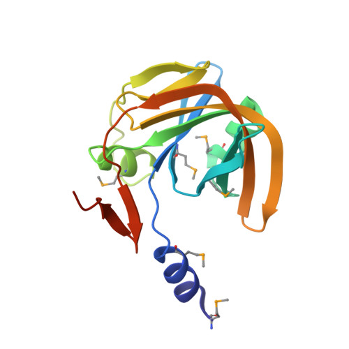
The catabolism of toxic phenols in the thermophilic organism Bacillus thermoglucosidasius A7 is initiated by a two-component enzyme system. The smaller flavin reductase PheA2 component catalyzes the NADH-dependent reduction of free FAD according to a ping-pong bisubstrate-biproduct mechanism. The reduced FAD is then used by the larger oxygenase component PheA1 to hydroxylate phenols to the corresponding catechols. We have determined the x-ray structure of PheA2 containing a bound FAD cofactor (2.2 A), which is the first structure of a member of this flavin reductase family. We have also determined the x-ray structure of reduced holo-PheA2 in complex with oxidized NAD (2.1 A). PheA2 is a single domain homodimeric protein with each FAD-containing subunit being organized around a six-stranded beta-sheet and a capping alpha-helix. The tightly bound FAD prosthetic group (K(d) = 10 nm) binds near the dimer interface, and the re face of the FAD isoalloxazine ring is fully exposed to solvent. The addition of NADH to crystalline PheA2 reduced the flavin cofactor, and the NAD product was bound in a wide solvent-accessible groove adopting an unusual folded conformation with ring stacking. This is the first observation of an enzyme that is very likely to react with a folded compact pyridine nucleotide. The PheA2 crystallographic models strongly suggest that reactive exogenous FAD substrate binds in the NADH cleft after release of NAD product. Nanoflow electrospray mass spectrometry data indeed showed that PheA2 is able to bind one FAD cofactor and one FAD substrate. In conclusion, the structural data provide evidence that PheA2 contains a dual binding cleft for NADH and FAD substrate, which alternate during catalysis.
Organizational Affiliation:
Department of Genetics and Microbiology, University of Pavia, via Abbiategrasso 207, 27100 Pavia, Italy. r.h.h.vandenheuvel@chem.uu.nl