1RYX

Crystal structure of hen serum transferrin in apo- form


Experimental Data Snapshot

  • Method: X-RAY DIFFRACTION
  • Resolution: 3.50 Å
  • R-Value Free: 0.341 
  • R-Value Work: 0.283 
  • R-Value Observed: 0.283 

wwPDB Validation   3D Report Full Report


This is version 1.4 of the entry. See complete history


Literature

Tertiary structural changes associated with iron binding and release in hen serum transferrin: a crystallographic and spectroscopic study

Thakurta, P.G.Choudhury, D.Dasgupta, R.Dattagupta, J.K.

(2004) Biochem Biophys Res Commun 316: 1124-1131

  • DOI: https://doi.org/10.1016/j.bbrc.2004.02.165
  • Primary Citation of Related Structures:  
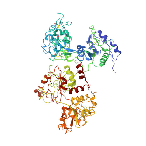
    1RYX

  • PubMed Abstract: 

    The iron binding and release of serum transferrin are pH-dependent and accompanied by a conformational change between the iron-bound (holo-) and iron-free (apo-) forms. We have determined the crystal structure of apo-hen serum transferrin (hAST) at 3.5A resolution, which is the first reported structure to date of any full molecule of an apo-serum transferrin and studied its pH-dependent iron release by UV-vis absorption and near UV-CD spectroscopy. The crystal structure of hAST shows that both the lobes adopt an open conformation and the relative orientations of the domains are different from those of apo-human serum transferrin and human apolactoferrin but similar to that of hen apo-ovotransferrin. Spectroscopic analysis reveals that in hen serum transferrin, release of the first iron starts at a pH approximately 6.5 and continues over a broad pH range (6.5-5.2). The complete release of the iron, however, occurs at pH approximately 4.0. The near UV-CD spectra show alterations in the microenvironment of the aromatic residues surrounding the iron-binding sites.


  • Organizational Affiliation

    Crystallography and Molecular Biology Division, Saha Institute of Nuclear Physics, 1/AF Bidhannagar, Kolkata 700 064, India.


Macromolecules
Find similar proteins by:  (by identity cutoff)  |  3D Structure
Entity ID: 1
MoleculeChains Sequence LengthOrganismDetailsImage
Ovotransferrin686Gallus gallusMutation(s): 0 
UniProt
Find proteins for P02789 (Gallus gallus)
Explore P02789 
Go to UniProtKB:  P02789
Entity Groups  
Sequence Clusters30% Identity50% Identity70% Identity90% Identity95% Identity100% Identity
UniProt GroupP02789
Sequence Annotations
Expand
  • Reference Sequence
Experimental Data & Validation

Experimental Data

  • Method: X-RAY DIFFRACTION
  • Resolution: 3.50 Å
  • R-Value Free: 0.341 
  • R-Value Work: 0.283 
  • R-Value Observed: 0.283 
  • Space Group: P 43 21 2
Unit Cell:
Length ( Å )Angle ( ˚ )
a = 90.295α = 90
b = 90.295β = 90
c = 177.666γ = 90
Software Package:
Software NamePurpose
CNSrefinement
DENZOdata reduction
SCALEPACKdata scaling
AMoREphasing

Structure Validation

View Full Validation Report


Entry History 

Deposition Data

Revision History  (Full details and data files)

  • Version 1.0: 2004-07-13
    Type: Initial release
  • Version 1.1: 2008-04-29
    Changes: Version format compliance
  • Version 1.2: 2011-07-13
    Changes: Version format compliance
  • Version 1.3: 2018-07-11
    Changes: Data collection
  • Version 1.4: 2023-10-25
    Changes: Data collection, Database references, Refinement description