1RXQ

YfiT from Bacillus subtilis is a probable metal-dependent hydrolase with an unusual four-helix bundle topology


Experimental Data Snapshot

  • Method: X-RAY DIFFRACTION
  • Resolution: 1.70 Å
  • R-Value Free: 0.217 
  • R-Value Work: 0.186 
  • R-Value Observed: 0.188 

wwPDB Validation   3D Report Full Report


This is version 1.2 of the entry. See complete history


Literature

YfiT from Bacillus subtilis Is a Probable Metal-Dependent Hydrolase with an Unusual Four-Helix Bundle Topology

Rajan, S.S.Yang, X.Shuvalova, L.Collart, F.Anderson, W.F.

(2004) Biochemistry 43: 15472-15479

  • DOI: https://doi.org/10.1021/bi048665r
  • Primary Citation of Related Structures:  
    1RXQ

  • PubMed Abstract: 

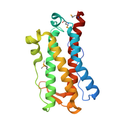
    YfiT, a 19-kDa polypeptide from Bacillus subtilis, belongs to a small sequence family with members predominantly from Gram positive bacteria. We have determined the crystal structure of YfiT in complex with Ni(2+) to a resolution of 1.7 A. YfiT exists as a dimer and binds Ni(2+) in a 1:1 stoichiometry. The protein has an unusual four-helix bundle topology and coordinates Ni(2+) in an octahedral geometry with three conserved histidines and three waters. Although there is no similarity in their overall structures, the coordination geometry of the metal and the residues that constitute the putative active site in YfiT are similar to those of metalloproteases such as thermolysin. Our structural analyses suggest that YfiT might function as a metal-dependent hydrolase.


  • Organizational Affiliation

    Molecular Pharmacology and Biological Chemistry, Feinberg School of Medicine, Northwestern University, Chicago, Illinois 60611, USA.


Macromolecules
Find similar proteins by:  (by identity cutoff)  |  3D Structure
Entity ID: 1
MoleculeChains Sequence LengthOrganismDetailsImage
yfiT
A, B, C, D
178Bacillus subtilisMutation(s): 4 
Gene Names: yfiT
UniProt
Find proteins for O31562 (Bacillus subtilis (strain 168))
Explore O31562 
Go to UniProtKB:  O31562
Entity Groups  
Sequence Clusters30% Identity50% Identity70% Identity90% Identity95% Identity100% Identity
UniProt GroupO31562
Sequence Annotations
Expand
  • Reference Sequence
Small Molecules
Ligands 6 Unique
IDChains Name / Formula / InChI Key2D Diagram3D Interactions
GLU
Query on GLU

Download Ideal Coordinates CCD File 
I [auth D]GLUTAMIC ACID
C5 H9 N O4
WHUUTDBJXJRKMK-VKHMYHEASA-N
THR
Query on THR

Download Ideal Coordinates CCD File 
L [auth D]THREONINE
C4 H9 N O3
AYFVYJQAPQTCCC-GBXIJSLDSA-N
SER
Query on SER

Download Ideal Coordinates CCD File 
J [auth D],
M [auth D]
SERINE
C3 H7 N O3
MTCFGRXMJLQNBG-REOHCLBHSA-N
ALA
Query on ALA

Download Ideal Coordinates CCD File 
K [auth D]ALANINE
C3 H7 N O2
QNAYBMKLOCPYGJ-REOHCLBHSA-N
GLY
Query on GLY

Download Ideal Coordinates CCD File 
N [auth D]GLYCINE
C2 H5 N O2
DHMQDGOQFOQNFH-UHFFFAOYSA-N
NI
Query on NI

Download Ideal Coordinates CCD File 
E [auth A],
F [auth B],
G [auth C],
H [auth D]
NICKEL (II) ION
Ni
VEQPNABPJHWNSG-UHFFFAOYSA-N
Modified Residues  1 Unique
IDChains TypeFormula2D DiagramParent
MSE
Query on MSE
A, B, C, D
L-PEPTIDE LINKINGC5 H11 N O2 SeMET
Experimental Data & Validation

Experimental Data

  • Method: X-RAY DIFFRACTION
  • Resolution: 1.70 Å
  • R-Value Free: 0.217 
  • R-Value Work: 0.186 
  • R-Value Observed: 0.188 
  • Space Group: P 1
Unit Cell:
Length ( Å )Angle ( ˚ )
a = 46.37α = 104.38
b = 50.42β = 90.61
c = 89.41γ = 112.05
Software Package:
Software NamePurpose
REFMACrefinement
MOSFLMdata reduction
CCP4data scaling
SOLVEphasing
RESOLVEphasing

Structure Validation

View Full Validation Report


Entry History 

Deposition Data

Revision History  (Full details and data files)

  • Version 1.0: 2004-07-20
    Type: Initial release
  • Version 1.1: 2008-04-29
    Changes: Version format compliance
  • Version 1.2: 2011-07-13
    Changes: Derived calculations, Version format compliance