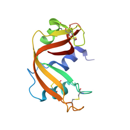
1ROB

STRUCTURE OF THE CRYSTALLINE COMPLEX OF CYTIDYLIC ACID (2'-CMP) WITH RIBONUCLEASE AT 1.6 ANGSTROMS RESOLUTION


Experimental Data Snapshot

  • Method: X-RAY DIFFRACTION
  • Resolution: 1.60 Å
  • R-Value Observed: 0.170 

wwPDB Validation   3D Report Full Report


This is version 1.2 of the entry. See complete history


Literature

Structure of the crystalline complex of cytidylic acid (2'-CMP) with ribonuclease at 1.6 A resolution. Conservation of solvent sites in RNase-A high-resolution structures.

Lisgarten, J.N.Gupta, V.Maes, D.Wyns, L.Zegers, I.Palmer, R.A.Dealwis, C.G.Aguilar, C.F.Hemmings, A.M.

(1993) Acta Crystallogr D Biol Crystallogr 49: 541-547

  • DOI: https://doi.org/10.1107/S090744499300719X
  • Primary Citation of Related Structures:  
    1ROB

  • PubMed Abstract: 

    The X-ray structure of the inhibitor complex of bovine ribonuclease A with cytidylic acid (2'-CMP) has been determined at 1.6 A resolution and refined by restrained least squares to R = 0.17 for 11 945 reflections. Binding of the inhibitor molecule to the protein is confirmed to be in the productive mode associated with enzyme activity. A study of conserved solvent sites amongst high-resolution structures in the same crystal form reveals a stabilizing water cluster between the N and C termini.


  • Organizational Affiliation

    Department of Ultrastructure, Institut voor Molekulaire Biologie, Vrije Universiteit Brussel, Sint Genesius Rode, Belgium.


Macromolecules
Find similar proteins by:  (by identity cutoff)  |  3D Structure
Entity ID: 1
MoleculeChains Sequence LengthOrganismDetailsImage
RIBONUCLEASE A124Bos taurusMutation(s): 0 
EC: 3.1.27.5
UniProt
Find proteins for P61823 (Bos taurus)
Explore P61823 
Go to UniProtKB:  P61823
Entity Groups  
Sequence Clusters30% Identity50% Identity70% Identity90% Identity95% Identity100% Identity
UniProt GroupP61823
Sequence Annotations
Expand
  • Reference Sequence
Small Molecules
Ligands 1 Unique
IDChains Name / Formula / InChI Key2D Diagram3D Interactions
C2P
Query on C2P

Download Ideal Coordinates CCD File 
B [auth A]CYTIDINE-2'-MONOPHOSPHATE
C9 H14 N3 O8 P
YQUAKORMLHPSLZ-XVFCMESISA-N
Binding Affinity Annotations 
IDSourceBinding Affinity
C2P BindingDB:  1ROB Ki: 7000 (nM) from 1 assay(s)
Experimental Data & Validation

Experimental Data

  • Method: X-RAY DIFFRACTION
  • Resolution: 1.60 Å
  • R-Value Observed: 0.170 
  • Space Group: P 1 21 1
Unit Cell:
Length ( Å )Angle ( ˚ )
a = 30.512α = 90
b = 38.242β = 106.1
c = 53.303γ = 90
Software Package:
Software NamePurpose
RESTRAINrefinement

Structure Validation

View Full Validation Report


Entry History 

Deposition Data

Revision History  (Full details and data files)

  • Version 1.0: 1994-01-31
    Type: Initial release
  • Version 1.1: 2008-03-24
    Changes: Version format compliance
  • Version 1.2: 2011-07-13
    Changes: Version format compliance