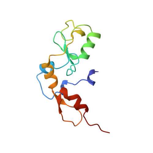
1RMD

RAG1 DIMERIZATION DOMAIN


Experimental Data Snapshot

  • Method: X-RAY DIFFRACTION
  • Resolution: 2.10 Å
  • R-Value Free: 0.257 
  • R-Value Work: 0.209 
  • R-Value Observed: 0.209 

wwPDB Validation   3D Report Full Report


This is version 1.3 of the entry. See complete history


Literature

Crystal structure of the RAG1 dimerization domain reveals multiple zinc-binding motifs including a novel zinc binuclear cluster.

Bellon, S.F.Rodgers, K.K.Schatz, D.G.Coleman, J.E.Steitz, T.A.

(1997) Nat Struct Biol 4: 586-591

  • DOI: https://doi.org/10.1038/nsb0797-586
  • Primary Citation of Related Structures:  
    1RMD

  • PubMed Abstract: 

    The crystal structure of the dimerization domain of the V(D)J recombination-activating protein, RAG1, was solved using zinc anomalous scattering. The structure reveals an unusual combination of multi-class zinc-binding motifs, including a zinc RING finger and a C2H2 zinc finger, that together from a single structural domain. The domain also contains a unique zinc binuclear cluster in place of a normally mononuclear zinc site in the RING finger. Together, four zinc ions help organize the entire domain, including the two helices that form the dimer interface.


  • Organizational Affiliation

    Department of Molecular Biophysics and Biochemistry, Yale University, New Haven, Connecticut 06520-8114, USA.


Macromolecules
Find similar proteins by:  (by identity cutoff)  |  3D Structure
Entity ID: 1
MoleculeChains Sequence LengthOrganismDetailsImage
RAG1116Mus musculusMutation(s): 0 
UniProt
Find proteins for P15919 (Mus musculus)
Explore P15919 
Go to UniProtKB:  P15919
Entity Groups  
Sequence Clusters30% Identity50% Identity70% Identity90% Identity95% Identity100% Identity
UniProt GroupP15919
Sequence Annotations
Expand
  • Reference Sequence
Experimental Data & Validation

Experimental Data

  • Method: X-RAY DIFFRACTION
  • Resolution: 2.10 Å
  • R-Value Free: 0.257 
  • R-Value Work: 0.209 
  • R-Value Observed: 0.209 
  • Space Group: P 32 2 1
Unit Cell:
Length ( Å )Angle ( ˚ )
a = 57.58α = 90
b = 57.58β = 90
c = 98.43γ = 120
Software Package:
Software NamePurpose
BAZFAZmodel building
X-PLORrefinement
DENZOdata reduction
SCALEPACKdata scaling
BAZFAZphasing

Structure Validation

View Full Validation Report


Entry History 

Deposition Data

Revision History  (Full details and data files)

  • Version 1.0: 1997-07-23
    Type: Initial release
  • Version 1.1: 2008-03-24
    Changes: Version format compliance
  • Version 1.2: 2011-07-13
    Changes: Version format compliance
  • Version 1.3: 2024-02-14
    Changes: Data collection, Database references, Derived calculations, Other, Structure summary