


Crystal structure of the complex of resistant strain of hiv-1 protease(v82a mutant) with ritonavir
Rezacova, P., Brynda, J., Sedlacek, J., Konvalinka, J., Fabry, M., Horejsi, M.To be published.
Experimental Data Snapshot
Entity ID: 1 | |||||
|---|---|---|---|---|---|
| Molecule | Chains | Sequence Length | Organism | Details | Image |
| protease RETROPEPSIN | 99 | Human immunodeficiency virus 1 | Mutation(s): 2 Gene Names: gag-pol EC: 3.4.23.16 | 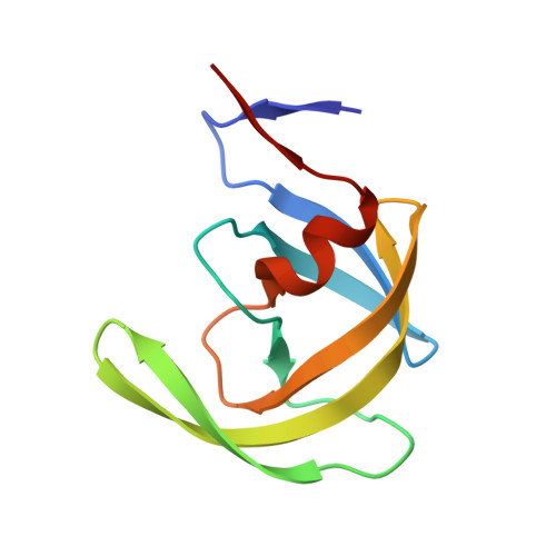 | |
UniProt | |||||
Find proteins for P03367 (Human immunodeficiency virus type 1 group M subtype B (isolate BRU/LAI)) Explore P03367 Go to UniProtKB: P03367 | |||||
Entity Groups | |||||
| Sequence Clusters | 30% Identity50% Identity70% Identity90% Identity95% Identity100% Identity | ||||
| UniProt Group | P03367 | ||||
Sequence AnnotationsExpand | |||||
| |||||
| Ligands 1 Unique | |||||
|---|---|---|---|---|---|
| ID | Chains | Name / Formula / InChI Key | 2D Diagram | 3D Interactions | |
| RIT Query on RIT | C [auth A] | RITONAVIR C37 H48 N6 O5 S2 NCDNCNXCDXHOMX-XGKFQTDJSA-N |  | ||
| Binding Affinity Annotations | |||
|---|---|---|---|
| ID | Source | Binding Affinity | |
| RIT | BindingDB: 1RL8 | Ki: min: 0.01, max: 109 (nM) from 36 assay(s) | |
| Kd: min: 0.6, max: 0.61 (nM) from 2 assay(s) | |||
| IC50: min: 67, max: 9850 (nM) from 2 assay(s) | |||
| EC50: 150 (nM) from 1 assay(s) | |||
| -TΔS: min: -1.02e+2, max: -3.01e+1 (kJ/mol) from 42 assay(s) | |||
| ΔH: min: -2.76e+1, max: 73.15 (kJ/mol) from 44 assay(s) | |||
| ΔG: min: -6.02e+1, max: -2.88e+1 (kJ/mol) from 42 assay(s) | |||
Entity ID: 2 | |||||
|---|---|---|---|---|---|
| ID | Chains | Name | Type/Class | 2D Diagram | 3D Interactions |
| PRD_001001 (RIT) Query on PRD_001001 | C [auth A] | RITONAVIR | Peptide-like / Inhibitor | | |
| Length ( Å ) | Angle ( ˚ ) |
|---|---|
| a = 61.414 | α = 90 |
| b = 61.414 | β = 90 |
| c = 80.816 | γ = 120 |
| Software Name | Purpose |
|---|---|
| CNS | refinement |
| SHELX | refinement |
| CNS | phasing |
RCSB PDB (citation) is hosted by
RCSB PDB is a member of the

