1RK0

Mhc Class I H-2Kb Heavy Chain Complexed With beta-2 Microglobulin and Herpes Simplex Virus Glycoprotein B peptide


Experimental Data Snapshot

  • Method: X-RAY DIFFRACTION
  • Resolution: 2.61 Å
  • R-Value Free: 0.238 
  • R-Value Work: 0.199 
  • R-Value Observed: 0.199 

wwPDB Validation   3D Report Full Report


This is version 2.0 of the entry. See complete history


Literature

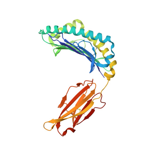
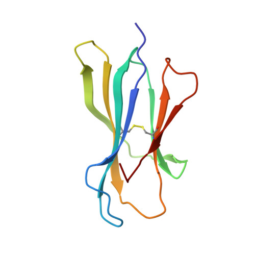
Structural Basis for the Restoration of TCR Recognition of an MHC Allelic Variant by Peptide Secondary Anchor Substitution

Miley, M.J.Messaoudi, I.Metzner, B.M.Wu, Y.Nikolich-Zugich, J.Fremont, D.H.

(2004) J Exp Med 200: 1445-1454

  • DOI: https://doi.org/10.1084/jem.20040217
  • Primary Citation of Related Structures:  
    1RJY, 1RJZ, 1RK0, 1RK1

  • PubMed Abstract: 

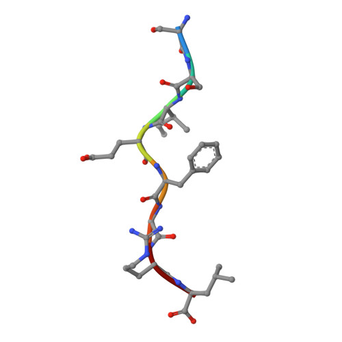
    Major histocompatibility complex (MHC) class I variants H-2K(b) and H-2K(bm8) differ primarily in the B pocket of the peptide-binding groove, which serves to sequester the P2 secondary anchor residue. This polymorphism determines resistance to lethal herpes simplex virus (HSV-1) infection by modulating T cell responses to the immunodominant glycoprotein B(498-505) epitope, HSV8. We studied the molecular basis of these effects and confirmed that T cell receptors raised against K(b)-HSV8 cannot recognize H-2K(bm8)-HSV8. However, substitution of Ser(P2) to Glu(P2) (peptide H2E) reversed T cell receptor (TCR) recognition; H-2K(bm8)-H2E was recognized whereas H-2K(b)-H2E was not. Insight into the structural basis of this discrimination was obtained by determining the crystal structures of all four MHC class I molecules in complex with bound peptide (pMHCs). Surprisingly, we find no concerted pMHC surface differences that can explain the differential TCR recognition. However, a correlation is apparent between the recognition data and the underlying peptide-binding groove chemistry of the B pocket, revealing that secondary anchor residues can profoundly affect TCR engagement through mechanisms distinct from the alteration of the resting state conformation of the pMHC surface.


  • Organizational Affiliation

    Department of Pathology and Immunology, Washington University School of Medicine, St. Louis, MO 63110, USA.


Macromolecules
Find similar proteins by:  (by identity cutoff)  |  3D Structure
Entity ID: 1
MoleculeChains Sequence LengthOrganismDetailsImage
H-2 class I histocompatibility antigen, K-B alpha chain274Mus musculusMutation(s): 0 
Gene Names: H2-K
UniProt
Find proteins for P01901 (Mus musculus)
Explore P01901 
Go to UniProtKB:  P01901
Entity Groups  
Sequence Clusters30% Identity50% Identity70% Identity90% Identity95% Identity100% Identity
UniProt GroupP01901
Sequence Annotations
Expand
  • Reference Sequence
Find similar proteins by:  (by identity cutoff)  |  3D Structure
Entity ID: 2
MoleculeChains Sequence LengthOrganismDetailsImage
Beta-2-microglobulin99Mus musculusMutation(s): 0 
Gene Names: B2M
UniProt & NIH Common Fund Data Resources
Find proteins for P01887 (Mus musculus)
Explore P01887 
Go to UniProtKB:  P01887
IMPC:  MGI:88127
Entity Groups  
Sequence Clusters30% Identity50% Identity70% Identity90% Identity95% Identity100% Identity
UniProt GroupP01887
Sequence Annotations
Expand
  • Reference Sequence

Find similar proteins by:  Sequence   |   3D Structure  

Entity ID: 3
MoleculeChains Sequence LengthOrganismDetailsImage
Glycoprotein BC [auth P]8N/AMutation(s): 0 
UniProt
Find proteins for P06436 (Human herpesvirus 1 (strain F))
Explore P06436 
Go to UniProtKB:  P06436
Entity Groups  
UniProt GroupP06436
Sequence Annotations
Expand
  • Reference Sequence
Small Molecules
Ligands 1 Unique
IDChains Name / Formula / InChI Key2D Diagram3D Interactions
NAG
Query on NAG

Download Ideal Coordinates CCD File 
D [auth A]2-acetamido-2-deoxy-beta-D-glucopyranose
C8 H15 N O6
OVRNDRQMDRJTHS-FMDGEEDCSA-N
Experimental Data & Validation

Experimental Data

  • Method: X-RAY DIFFRACTION
  • Resolution: 2.61 Å
  • R-Value Free: 0.238 
  • R-Value Work: 0.199 
  • R-Value Observed: 0.199 
  • Space Group: P 21 21 2
Unit Cell:
Length ( Å )Angle ( ˚ )
a = 134.89α = 90
b = 90.222β = 90
c = 45.451γ = 90
Software Package:
Software NamePurpose
CNSrefinement
HKL-2000data reduction
SCALEPACKdata scaling
CNSphasing

Structure Validation

View Full Validation Report


Entry History 

Deposition Data

Revision History  (Full details and data files)

  • Version 1.0: 2004-12-14
    Type: Initial release
  • Version 1.1: 2008-04-29
    Changes: Version format compliance
  • Version 1.2: 2011-07-13
    Changes: Version format compliance
  • Version 2.0: 2020-07-29
    Type: Remediation
    Reason: Carbohydrate remediation
    Changes: Advisory, Atomic model, Data collection, Derived calculations, Non-polymer description, Structure summary