1RJ8

The crystal structure of TNF family member EDA-A2


Experimental Data Snapshot

  • Method: X-RAY DIFFRACTION
  • Resolution: 2.23 Å
  • R-Value Free: 0.241 
  • R-Value Work: 0.200 
  • R-Value Observed: 0.204 

wwPDB Validation   3D Report Full Report


This is version 1.6 of the entry. See complete history


Literature

The Crystal Structure of EDA-A1 and EDA-A2: splice variants with distinct receptor specificity

Hymowitz, S.G.Compaan, D.M.Yan, M.Ackerly, H.Dixit, V.M.Starovasnik, M.A.de Vos, A.M.

(2003) Structure 11: 1513-1520

  • DOI: https://doi.org/10.1016/j.str.2003.11.009
  • Primary Citation of Related Structures:  
    1RJ7, 1RJ8

  • PubMed Abstract: 

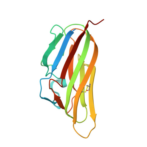
    EDA is a tumor necrosis factor family member involved in ectodermal development. Splice variants EDA-A1 and EDA-A2 differ only by the presence of Glu 308 and Val 309 in the expected receptor binding region of EDA-A1 but not EDA-A2. This two amino acid difference functions as a switch controlling receptor specificity. EDA-A1 binds only to EDAR, while EDA-A2 is specific for XEDAR. In order to understand the structural basis of this switch, we determined the X-ray crystal structures of the TNF domain of both EDA-A1 and EDA-A2 at 2.3 A and 2.2 A, respectively. While the backbone conformation around the splice difference is similar in both isoforms, the conformation of the following loop, the surface charge, and the shape of the expected receptor binding site differ significantly.


  • Organizational Affiliation

    Department of Protein Engineering, Genentech, Inc., 1 DNA Way, South San Francisco, CA 94080, USA. hymowitz@gene.com


Macromolecules
Find similar proteins by:  (by identity cutoff)  |  3D Structure
Entity ID: 1
MoleculeChains Sequence LengthOrganismDetailsImage
ectodysplasin-A isoform EDA-A2164Homo sapiensMutation(s): 0 
Gene Names: EDA genesplice form EDA-A2
UniProt & NIH Common Fund Data Resources
Find proteins for Q92838 (Homo sapiens)
Explore Q92838 
Go to UniProtKB:  Q92838
PHAROS:  Q92838
GTEx:  ENSG00000158813 
Entity Groups  
Sequence Clusters30% Identity50% Identity70% Identity90% Identity95% Identity100% Identity
UniProt GroupQ92838
Sequence Annotations
Expand
  • Reference Sequence
Experimental Data & Validation

Experimental Data

  • Method: X-RAY DIFFRACTION
  • Resolution: 2.23 Å
  • R-Value Free: 0.241 
  • R-Value Work: 0.200 
  • R-Value Observed: 0.204 
  • Space Group: P 1 21 1
Unit Cell:
Length ( Å )Angle ( ˚ )
a = 50.723α = 90
b = 161.337β = 105.24
c = 51.023γ = 90
Software Package:
Software NamePurpose
REFMACrefinement
MAR345data collection
XDSdata scaling
AMoREphasing

Structure Validation

View Full Validation Report


Entry History 

Deposition Data

Revision History  (Full details and data files)

  • Version 1.0: 2003-12-09
    Type: Initial release
  • Version 1.1: 2008-04-29
    Changes: Version format compliance
  • Version 1.2: 2011-07-13
    Changes: Advisory, Version format compliance
  • Version 1.3: 2017-10-11
    Changes: Refinement description
  • Version 1.4: 2018-01-31
    Changes: Experimental preparation
  • Version 1.5: 2018-04-04
    Changes: Data collection, Database references
  • Version 1.6: 2024-04-03
    Changes: Data collection, Database references, Refinement description