1RIS

CRYSTAL STRUCTURE OF THE RIBOSOMAL PROTEIN S6 FROM THERMUS THERMOPHILUS


Experimental Data Snapshot

  • Method: X-RAY DIFFRACTION
  • Resolution: 2.00 Å
  • R-Value Work: 0.183 
  • R-Value Observed: 0.183 

wwPDB Validation   3D Report Full Report


This is version 1.4 of the entry. See complete history


Literature

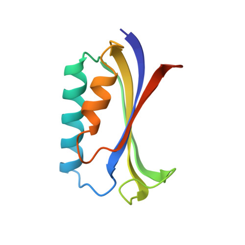
Crystal structure of the ribosomal protein S6 from Thermus thermophilus.

Lindahl, M.Svensson, L.A.Liljas, A.Sedelnikova, S.E.Eliseikina, I.A.Fomenkova, N.P.Nevskaya, N.Nikonov, S.V.Garber, M.B.Muranova, T.A.Rykonova, A.I.Amons, R.

(1994) EMBO J 13: 1249-1254

  • DOI: https://doi.org/10.2210/pdb1ris/pdb
  • Primary Citation of Related Structures:  
    1RIS

  • PubMed Abstract: 

    The amino acid sequence and crystal structure of the ribosomal protein S6 from the small ribosomal subunit of Thermus thermophilus have been determined. S6 is a small protein with 101 amino acid residues. The 3D structure, which was determined to 2.0 A resolution, consists of a four-stranded anti-parallel beta-sheet with two alpha-helices packed on one side. Similar folding patterns have been observed for other ribosomal proteins and may suggest an original RNA-interacting motif. Related topologies are also found in several other nucleic acid-interacting proteins and based on the assumption that the structure of the ribosome was established early in the molecular evolution, the possibility that an ancestral RNA-interacting motif in ribosomal proteins is the evolutionary origin for the nucleic acid-interacting domain in large classes of ribonucleic acid binding proteins should be considered.


  • Organizational Affiliation

    Chemical Center, University of Lund, Sweden.


Macromolecules
Find similar proteins by:  (by identity cutoff)  |  3D Structure
Entity ID: 1
MoleculeChains Sequence LengthOrganismDetailsImage
RIBOSOMAL PROTEIN S6101Thermus thermophilusMutation(s): 0 
UniProt
Find proteins for P23370 (Thermus thermophilus)
Explore P23370 
Go to UniProtKB:  P23370
Entity Groups  
Sequence Clusters30% Identity50% Identity70% Identity90% Identity95% Identity100% Identity
UniProt GroupP23370
Sequence Annotations
Expand
  • Reference Sequence
Experimental Data & Validation

Experimental Data

  • Method: X-RAY DIFFRACTION
  • Resolution: 2.00 Å
  • R-Value Work: 0.183 
  • R-Value Observed: 0.183 
  • Space Group: C 2 2 2
Unit Cell:
Length ( Å )Angle ( ˚ )
a = 106.81α = 90
b = 52.72β = 90
c = 41.12γ = 90
Software Package:
Software NamePurpose
X-PLORmodel building
X-PLORrefinement
X-PLORphasing

Structure Validation

View Full Validation Report


Entry History 

Revision History  (Full details and data files)

  • Version 1.0: 1994-09-30
    Type: Initial release
  • Version 1.1: 2008-03-24
    Changes: Version format compliance
  • Version 1.2: 2011-07-13
    Changes: Version format compliance
  • Version 1.3: 2017-11-29
    Changes: Derived calculations, Other
  • Version 1.4: 2024-02-14
    Changes: Data collection, Database references