1RE4

Crystal Structure of Fragment D of BbetaD398A Fibrinogen


Experimental Data Snapshot

  • Method: X-RAY DIFFRACTION
  • Resolution: 2.70 Å
  • R-Value Free: 0.271 
  • R-Value Work: 0.220 
  • R-Value Observed: 0.220 

wwPDB Validation   3D Report Full Report


Ligand Structure Quality Assessment 


This is version 1.5 of the entry. See complete history


Literature

BbetaGlu397 and BbetaAsp398 but not BbetaAsp432 are required for "B:b" interactions.

Kostelansky, M.S.Bolliger-Stucki, B.Betts, L.Gorkun, O.V.Lord, S.T.

(2004) Biochemistry 43: 2465-2474

  • DOI: https://doi.org/10.1021/bi035996f
  • Primary Citation of Related Structures:  
    1RE3, 1RE4

  • PubMed Abstract: 

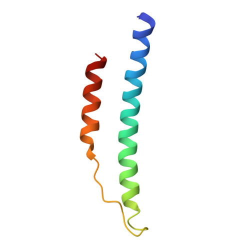
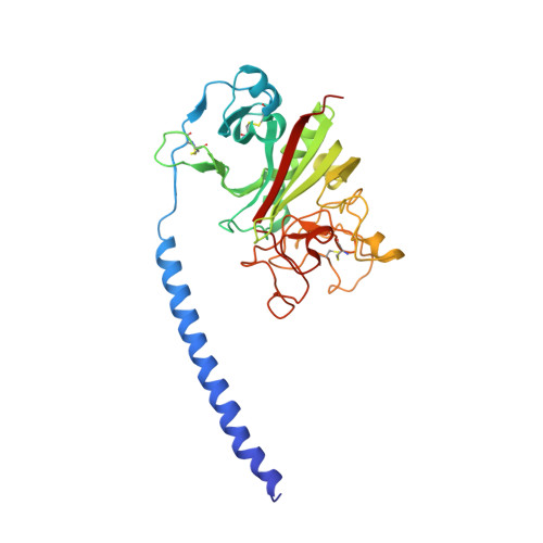
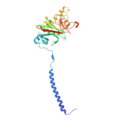
    We synthesized three fibrinogen variants, BbetaE397A, BbetaD398A, and BbetaD432A, with substitutions at positions identified in crystallographic studies as critical for binding the "B" peptide, Gly-His-Arg-Pro-amide (GHRPam), to the "b" polymerization site. We examined thrombin- and batroxobin-catalyzed polymerization by turbidity measurements and found that BbetaE397A and BbetaD398A were impaired while BbetaD432A was normal. Changes in polymerization as a function of calcium were similar for variant and normal fibrinogens. We determined crystal structures of fragment D from the variant BbetaD398A in the absence and presence of GHRPam. In the absence of peptide, the structure showed that the alanine substitution altered only specific local interactions, as alignment of the variant structure with the analogous normal structure resulted in an RMSD of 0.53 A over all atoms. The structure also showed reduced occupancy of the beta2 calcium-binding site that includes the side chain carbonyl of BbetaD398, suggesting that calcium was not bound at this site in our polymerization studies. In the presence of peptide, the structure showed that GHRPam was not bound in the "b" site and the conformational changes associated with peptide binding to normal fragment D did not occur. This structure also showed GHRPam bound in the "a" polymerization site, although in two different conformations. Calcium binding was associated with only one of these conformations, suggesting that calcium binding to the gamma2-site and an alternative peptide conformation were induced by crystal packing. We conclude that BbetaE397 and BbetaD398 are essential for the "B:b" interaction, while BbetaD432 is not.


  • Organizational Affiliation

    Department of Chemistry, University of North Carolina, Chapel Hill, North Carolina 27599-7525, USA.


Macromolecules
Find similar proteins by:  (by identity cutoff)  |  3D Structure
Entity ID: 1
MoleculeChains Sequence LengthOrganismDetailsImage
Fibrinogen alpha/alpha-E chain
A, D
66Homo sapiensMutation(s): 0 
Gene Names: FGA
UniProt & NIH Common Fund Data Resources
Find proteins for P02671 (Homo sapiens)
Explore P02671 
Go to UniProtKB:  P02671
PHAROS:  P02671
GTEx:  ENSG00000171560 
Entity Groups  
Sequence Clusters30% Identity50% Identity70% Identity90% Identity95% Identity100% Identity
UniProt GroupP02671
Sequence Annotations
Expand
  • Reference Sequence
Find similar proteins by:  (by identity cutoff)  |  3D Structure
Entity ID: 2
MoleculeChains Sequence LengthOrganismDetailsImage
Fibrinogen beta chain
B, E
313Homo sapiensMutation(s): 1 
Gene Names: FGB
UniProt & NIH Common Fund Data Resources
Find proteins for P02675 (Homo sapiens)
Explore P02675 
Go to UniProtKB:  P02675
PHAROS:  P02675
GTEx:  ENSG00000171564 
Entity Groups  
Sequence Clusters30% Identity50% Identity70% Identity90% Identity95% Identity100% Identity
UniProt GroupP02675
Sequence Annotations
Expand
  • Reference Sequence
Find similar proteins by:  (by identity cutoff)  |  3D Structure
Entity ID: 3
MoleculeChains Sequence LengthOrganismDetailsImage
Fibrinogen gamma chain
C, F
311Homo sapiensMutation(s): 0 
Gene Names: FGG
UniProt & NIH Common Fund Data Resources
Find proteins for P02679 (Homo sapiens)
Explore P02679 
Go to UniProtKB:  P02679
PHAROS:  P02679
GTEx:  ENSG00000171557 
Entity Groups  
Sequence Clusters30% Identity50% Identity70% Identity90% Identity95% Identity100% Identity
UniProt GroupP02679
Sequence Annotations
Expand
  • Reference Sequence
Experimental Data & Validation

Experimental Data

  • Method: X-RAY DIFFRACTION
  • Resolution: 2.70 Å
  • R-Value Free: 0.271 
  • R-Value Work: 0.220 
  • R-Value Observed: 0.220 
  • Space Group: P 21 21 21
Unit Cell:
Length ( Å )Angle ( ˚ )
a = 88.538α = 90
b = 94.487β = 90
c = 227.088γ = 90
Software Package:
Software NamePurpose
CNSrefinement
HKL-2000data reduction
SCALEPACKdata scaling
CNSphasing

Structure Validation

View Full Validation Report


Ligand Structure Quality Assessment 


Entry History 

Deposition Data

Revision History  (Full details and data files)

  • Version 1.0: 2004-05-25
    Type: Initial release
  • Version 1.1: 2008-04-29
    Changes: Version format compliance
  • Version 1.2: 2011-07-13
    Changes: Non-polymer description, Version format compliance
  • Version 1.3: 2020-07-29
    Type: Remediation
    Reason: Carbohydrate remediation
    Changes: Data collection, Derived calculations, Structure summary
  • Version 1.4: 2021-10-27
    Changes: Database references, Structure summary
  • Version 1.5: 2023-08-23
    Changes: Data collection, Refinement description