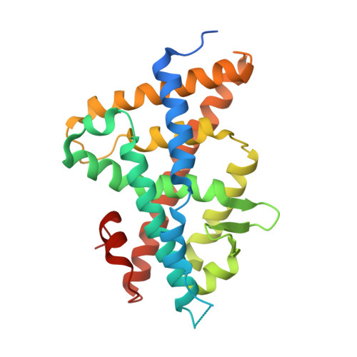
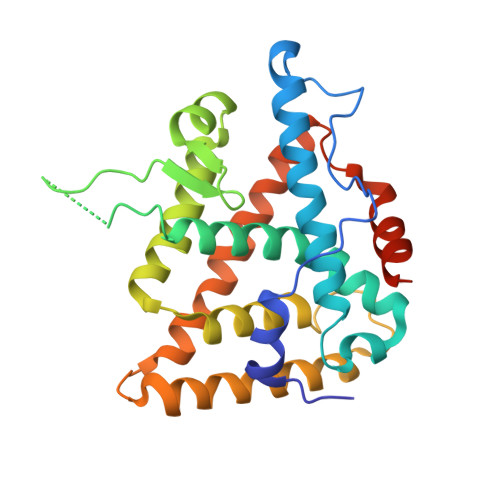
1R1K

Crystal structure of the ligand-binding domains of the heterodimer EcR/USP bound to ponasterone A


Experimental Data Snapshot

  • Method: X-RAY DIFFRACTION
  • Resolution: 2.90 Å
  • R-Value Free: 0.304 
  • R-Value Work: 0.243 
  • R-Value Observed: 0.243 

wwPDB Validation   3D Report Full Report


Ligand Structure Quality Assessment 


This is version 1.3 of the entry. See complete history


Literature

Structural adaptability in the ligand-binding pocket of the ecdysone hormone receptor.

Billas, I.M.L.Iwema, T.Garnier, J.M.Mitschler, A.Rochel, N.Moras, D.

(2003) Nature 426: 91-96

  • DOI: https://doi.org/10.1038/nature02112
  • Primary Citation of Related Structures:  
    1R1K, 1R20

  • PubMed Abstract: 

    The ecdysteroid hormones coordinate the major stages of insect development, notably moulting and metamorphosis, by binding to the ecdysone receptor (EcR); a ligand-inducible nuclear transcription factor. To bind either ligand or DNA, EcR must form a heterodimer with ultraspiracle (USP), the homologue of retinoid-X receptor. Here we report the crystal structures of the ligand-binding domains of the moth Heliothis virescens EcR-USP heterodimer in complex with the ecdysteroid ponasterone A and with a non-steroidal, lepidopteran-specific agonist BYI06830 used in agrochemical pest control. The two structures of EcR-USP emphasize the universality of heterodimerization as a general mechanism common to both vertebrates and invertebrates. Comparison of the EcR structures in complex with steroidal and non-steroidal ligands reveals radically different and only partially overlapping ligand-binding pockets that could not be predicted by molecular modelling and docking studies. These findings offer new perspectives for the design of insect-specific, environmentally safe insecticides. The concept of a ligand-dependent binding pocket in EcR provides an insight into the moulding of nuclear receptors to their ligand, and has potential applications for human nuclear receptors.


  • Organizational Affiliation

    Département de Biologie et de Génomique Structurales, IGBMC, CNRS/INSERM/Université Louis Pasteur, Parc d'Innovation BP10142, 67404 Illkirch cedex, France.


Macromolecules
Find similar proteins by:  (by identity cutoff)  |  3D Structure
Entity ID: 1
MoleculeChains Sequence LengthOrganismDetailsImage
Ecdysone receptorA [auth D]266Heliothis virescensMutation(s): 0 
Gene Names: ECR OR NR1H1
UniProt
Find proteins for O18473 (Heliothis virescens)
Explore O18473 
Go to UniProtKB:  O18473
Entity Groups  
Sequence Clusters30% Identity50% Identity70% Identity90% Identity95% Identity100% Identity
UniProt GroupO18473
Sequence Annotations
Expand
  • Reference Sequence
Find similar proteins by:  (by identity cutoff)  |  3D Structure
Entity ID: 2
MoleculeChains Sequence LengthOrganismDetailsImage
ULTRASPIRACLE PROTEINB [auth A]263Heliothis virescensMutation(s): 0 
UniProt
Find proteins for Q7SIF6 (Heliothis virescens)
Explore Q7SIF6 
Go to UniProtKB:  Q7SIF6
Entity Groups  
Sequence Clusters30% Identity50% Identity70% Identity90% Identity95% Identity100% Identity
UniProt GroupQ7SIF6
Sequence Annotations
Expand
  • Reference Sequence
Small Molecules
Ligands 2 Unique
IDChains Name / Formula / InChI Key2D Diagram3D Interactions
EPH
Query on EPH

Download Ideal Coordinates CCD File 
D [auth A]L-ALPHA-PHOSPHATIDYL-BETA-OLEOYL-GAMMA-PALMITOYL-PHOSPHATIDYLETHANOLAMINE
C39 H68 N O8 P
MABRTXOVHMDVAT-AAEGOEIASA-N
P1A
Query on P1A

Download Ideal Coordinates CCD File 
C [auth D]2,3,14,20,22-PENTAHYDROXYCHOLEST-7-EN-6-ONE
C27 H44 O6
PJYYBCXMCWDUAZ-JJJZTNILSA-N
Experimental Data & Validation

Experimental Data

  • Method: X-RAY DIFFRACTION
  • Resolution: 2.90 Å
  • R-Value Free: 0.304 
  • R-Value Work: 0.243 
  • R-Value Observed: 0.243 
  • Space Group: P 31 2 1
Unit Cell:
Length ( Å )Angle ( ˚ )
a = 57.401α = 90
b = 57.401β = 90
c = 302.696γ = 120
Software Package:
Software NamePurpose
HKL-2000data collection
SCALEPACKdata scaling
EPMRphasing
CNSrefinement
HKL-2000data reduction

Structure Validation

View Full Validation Report


Ligand Structure Quality Assessment 


Entry History 

Deposition Data

Revision History  (Full details and data files)

  • Version 1.0: 2003-11-18
    Type: Initial release
  • Version 1.1: 2008-04-29
    Changes: Version format compliance
  • Version 1.2: 2011-07-13
    Changes: Version format compliance
  • Version 1.3: 2023-08-23
    Changes: Data collection, Database references, Derived calculations, Refinement description, Structure summary