1QYY

Crystal Structure of N-Terminal Domain of Human Platelet Receptor Glycoprotein Ib-alpha at 2.8 Angstrom Resolution


Experimental Data Snapshot

  • Method: X-RAY DIFFRACTION
  • Resolution: 2.80 Å
  • R-Value Free: 0.282 
  • R-Value Work: 0.219 
  • R-Value Observed: 0.223 

wwPDB Validation   3D Report Full Report


Ligand Structure Quality Assessment 


This is version 2.1 of the entry. See complete history


Literature

Platinum-induced space-group transformation in crystals of the platelet glycoprotein Ib alpha N-terminal domain.

Varughese, K.I.Ruggeri, Z.M.Celikel, R.

(2004) Acta Crystallogr D Biol Crystallogr 60: 405-411

  • DOI: https://doi.org/10.1107/S0907444903026805
  • Primary Citation of Related Structures:  
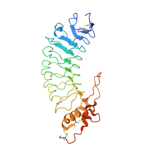
    1QYY

  • PubMed Abstract: 

    The interaction between platelet glycoprotein (GP) Ib alpha and von Willebrand factor (VWF) is essential for thrombus formation, leading to the arrest of bleeding. The N-terminal domain of GP Ib alpha, which contains the binding sites for VWF and alpha-thrombin, crystallized in the tetragonal space group P4(3) with one molecule in the asymmetric unit. When the crystals were treated with platinum, the crystals changed their symmetry from tetragonal to monoclinic P2(1) with two molecules in the asymmetric unit. The structure of the monoclinic form was solved using two-wavelength platinum anomalous dispersion data. The tetragonal crystal structure was subsequently solved using molecular-replacement techniques using the monoclinic structure as the search model and was refined with 1.7 A resolution data.


  • Organizational Affiliation

    Department of Molecular and Experimental Medicine, The Scripps Research Institute, La Jolla, CA 92037, USA. kiv@scripps.edu


Macromolecules
Find similar proteins by:  (by identity cutoff)  |  3D Structure
Entity ID: 1
MoleculeChains Sequence LengthOrganismDetailsImage
Platelet glycoprotein Ib alpha chainA,
B [auth G]
290Homo sapiensMutation(s): 1 
Gene Names: GP1BA
UniProt & NIH Common Fund Data Resources
Find proteins for P07359 (Homo sapiens)
Explore P07359 
Go to UniProtKB:  P07359
PHAROS:  P07359
GTEx:  ENSG00000185245 
Entity Groups  
Sequence Clusters30% Identity50% Identity70% Identity90% Identity95% Identity100% Identity
UniProt GroupP07359
Sequence Annotations
Expand
  • Reference Sequence
Oligosaccharides

Help

Entity ID: 2
MoleculeChains Length2D Diagram Glycosylation3D Interactions
alpha-D-mannopyranose-(1-3)-alpha-D-mannopyranose-(1-4)-2-acetamido-2-deoxy-beta-D-glucopyranose-(1-4)-2-acetamido-2-deoxy-beta-D-glucopyranoseC [auth B]4N-Glycosylation
Glycosylation Resources
GlyTouCan:  G48068RF
GlyCosmos:  G48068RF
GlyGen:  G48068RF
Experimental Data & Validation

Experimental Data

  • Method: X-RAY DIFFRACTION
  • Resolution: 2.80 Å
  • R-Value Free: 0.282 
  • R-Value Work: 0.219 
  • R-Value Observed: 0.223 
  • Space Group: P 1 21 1
Unit Cell:
Length ( Å )Angle ( ˚ )
a = 51.61α = 90
b = 113.82β = 95.21
c = 56.21γ = 90
Software Package:
Software NamePurpose
MOSFLMdata reduction
SCALEPACKdata scaling
SOLVEphasing
CNSrefinement

Structure Validation

View Full Validation Report


Ligand Structure Quality Assessment 


Entry History 

Deposition Data

Revision History  (Full details and data files)

  • Version 1.0: 2004-03-02
    Type: Initial release
  • Version 1.1: 2008-04-29
    Changes: Version format compliance
  • Version 1.2: 2011-07-13
    Changes: Non-polymer description, Version format compliance
  • Version 2.0: 2020-07-29
    Type: Remediation
    Reason: Carbohydrate remediation
    Changes: Advisory, Atomic model, Data collection, Derived calculations, Structure summary
  • Version 2.1: 2021-10-27
    Changes: Database references, Structure summary