1QVI

Crystal structure of scallop myosin S1 in the pre-power stroke state to 2.6 Angstrom resolution: flexibility and function in the head


Experimental Data Snapshot

  • Method: X-RAY DIFFRACTION
  • Resolution: 2.54 Å
  • R-Value Free: 0.266 
  • R-Value Work: 0.212 
  • R-Value Observed: 0.212 

wwPDB Validation   3D Report Full Report


Ligand Structure Quality Assessment 


This is version 1.3 of the entry. See complete history


Literature

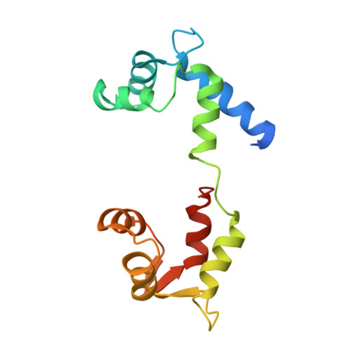
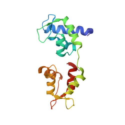
Crystal structure of scallop Myosin s1 in the pre-power stroke state to 2.6 a resolution: flexibility and function in the head.

Gourinath, S.Himmel, D.M.Brown, J.H.Reshetnikova, L.Szent-Gyorgyi, A.G.Cohen, C.

(2003) Structure 11: 1621-1627

  • DOI: https://doi.org/10.1016/j.str.2003.10.013
  • Primary Citation of Related Structures:  
    1QVI

  • PubMed Abstract: 

    We have extended the X-ray structure determination of the complete scallop myosin head in the pre-power stroke state to 2.6 A resolution, allowing an atomic comparison of the three major (weak actin binding) states of various myosins. We can now account for conformational differences observed in crystal structures in the so-called "pliant region" at the motor domain-lever arm junction between scallop and vertebrate smooth muscle myosins. A hinge, which may contribute to the compliance of the myosin crossbridge, has also been identified for the first time within the regulatory light-chain domain of the lever arm. Analysis of temperature factors of key joints of the motor domain, especially the SH1 helix, provides crystallographic evidence for the existence of the "internally uncoupled" state in diverse isoforms. The agreement between structural and solution studies reinforces the view that the unwinding of the SH1 helix is a part of the cross-bridge cycle in many myosins.


  • Organizational Affiliation

    Rosenstiel Basic Medical Sciences Research Center, Brandeis University, Waltham, MA 02454, USA.


Macromolecules
Find similar proteins by:  (by identity cutoff)  |  3D Structure
Entity ID: 1
MoleculeChains Sequence LengthOrganismDetailsImage
Myosin heavy chain, striated muscle840Argopecten irradiansMutation(s): 0 
UniProt
Find proteins for P24733 (Argopecten irradians)
Explore P24733 
Go to UniProtKB:  P24733
Entity Groups  
Sequence Clusters30% Identity50% Identity70% Identity90% Identity95% Identity100% Identity
UniProt GroupP24733
Sequence Annotations
Expand
  • Reference Sequence
Find similar proteins by:  (by identity cutoff)  |  3D Structure
Entity ID: 2
MoleculeChains Sequence LengthOrganismDetailsImage
Myosin regulatory light chain, striated adductor muscleB [auth Y]156Argopecten irradiansMutation(s): 0 
UniProt
Find proteins for P13543 (Argopecten irradians)
Explore P13543 
Go to UniProtKB:  P13543
Entity Groups  
Sequence Clusters30% Identity50% Identity70% Identity90% Identity95% Identity100% Identity
UniProt GroupP13543
Sequence Annotations
Expand
  • Reference Sequence
Find similar proteins by:  (by identity cutoff)  |  3D Structure
Entity ID: 3
MoleculeChains Sequence LengthOrganismDetailsImage
Myosin essential light chain, striated adductor muscleC [auth Z]156Argopecten irradiansMutation(s): 0 
UniProt
Find proteins for P07291 (Argopecten irradians)
Explore P07291 
Go to UniProtKB:  P07291
Entity Groups  
Sequence Clusters30% Identity50% Identity70% Identity90% Identity95% Identity100% Identity
UniProt GroupP07291
Sequence Annotations
Expand
  • Reference Sequence
Experimental Data & Validation

Experimental Data

  • Method: X-RAY DIFFRACTION
  • Resolution: 2.54 Å
  • R-Value Free: 0.266 
  • R-Value Work: 0.212 
  • R-Value Observed: 0.212 
  • Space Group: P 1 21 1
Unit Cell:
Length ( Å )Angle ( ˚ )
a = 51.375α = 90
b = 285.623β = 114.5
c = 59.831γ = 90
Software Package:
Software NamePurpose
CNSrefinement
DENZOdata reduction
SCALEPACKdata scaling
AMoREphasing

Structure Validation

View Full Validation Report


Ligand Structure Quality Assessment 


Entry History 

Deposition Data

Revision History  (Full details and data files)

  • Version 1.0: 2003-12-16
    Type: Initial release
  • Version 1.1: 2008-04-29
    Changes: Version format compliance
  • Version 1.2: 2011-07-13
    Changes: Version format compliance
  • Version 1.3: 2023-08-16
    Changes: Data collection, Database references, Derived calculations, Refinement description