1QU7

FOUR HELICAL-BUNDLE STRUCTURE OF THE CYTOPLASMIC DOMAIN OF A SERINE CHEMOTAXIS RECEPTOR


Experimental Data Snapshot

  • Method: X-RAY DIFFRACTION
  • Resolution: 2.60 Å
  • R-Value Free: 0.276 
  • R-Value Work: 0.243 
  • R-Value Observed: 0.243 

wwPDB Validation   3D Report Full Report


This is version 1.5 of the entry. See complete history


Literature

Four-helical-bundle structure of the cytoplasmic domain of a serine chemotaxis receptor.

Kim, K.K.Yokota, H.Kim, S.H.

(1999) Nature 400: 787-792

  • DOI: https://doi.org/10.1038/23512
  • Primary Citation of Related Structures:  
    1QU7

  • PubMed Abstract: 

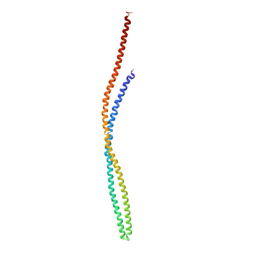
    The bacterial chemotaxis receptors are transmembrane receptors with a simple signalling pathway which has elements relevant to the general understanding of signal recognition and transduction across membranes, how signals are relayed between molecules in a pathway, and how adaptation to a persistent signal is achieved. In contrast to many mammalian receptors which signal by oligomerizing upon ligand binding, the chemotaxis receptors are dimeric even in the absence of their ligands, and their signalling does not depend on a monomer-dimer equilibrium. Bacterial chemotaxis receptors are composed of a ligand-binding domain, a transmembrane domain consisting of two helices TM1 and TM2, and a cytoplasmic domain. All known bacterial chemotaxis receptors have a highly conserved cytoplasmic domain, which unites signals from different ligand domains into a single signalling pathway to flagella motors. Here we report the crystal structure of the cytoplasmic domain of a serine chemotaxis receptor of Escherichia coli, which reveals a 200 A-long coiled-coil of two antiparallel helices connected by a 'U-turn'. Two of these domains form a long, supercoiled, four-helical bundle in the cytoplasmic portion of the receptor.


  • Organizational Affiliation

    Department of Chemistry, University of California, Berkeley 94720-5230, USA.


Macromolecules
Find similar proteins by:  (by identity cutoff)  |  3D Structure
Entity ID: 1
MoleculeChains Sequence LengthOrganismDetailsImage
METHYL-ACCEPTING CHEMOTAXIS PROTEIN I
A, B
227Escherichia coliMutation(s): 2 
UniProt
Find proteins for P02942 (Escherichia coli (strain K12))
Explore P02942 
Go to UniProtKB:  P02942
Entity Groups  
Sequence Clusters30% Identity50% Identity70% Identity90% Identity95% Identity100% Identity
UniProt GroupP02942
Sequence Annotations
Expand
  • Reference Sequence
Experimental Data & Validation

Experimental Data

  • Method: X-RAY DIFFRACTION
  • Resolution: 2.60 Å
  • R-Value Free: 0.276 
  • R-Value Work: 0.243 
  • R-Value Observed: 0.243 
  • Space Group: P 3 2 1
Unit Cell:
Length ( Å )Angle ( ˚ )
a = 132.44α = 90
b = 132.44β = 90
c = 134.59γ = 120
Software Package:
Software NamePurpose
MLPHAREphasing
CNSrefinement
MAR345data collection
SCALEPACKdata scaling

Structure Validation

View Full Validation Report


Entry History 

Deposition Data

Revision History  (Full details and data files)

  • Version 1.0: 2000-07-12
    Type: Initial release
  • Version 1.1: 2008-04-27
    Changes: Version format compliance
  • Version 1.2: 2011-07-13
    Changes: Version format compliance
  • Version 1.3: 2017-10-04
    Changes: Refinement description
  • Version 1.4: 2021-11-03
    Changes: Database references
  • Version 1.5: 2024-02-14
    Changes: Data collection