1QTS

CRYSTAL STRUCTURE OF THE AP-2 CLATHRIN ADAPTOR ALPHA-APPENDAGE


Experimental Data Snapshot

  • Method: X-RAY DIFFRACTION
  • Resolution: 1.40 Å
  • R-Value Free: 0.209 
  • R-Value Work: 0.178 

wwPDB Validation   3D Report Full Report


This is version 1.3 of the entry. See complete history


Literature

Crystal structure of the alpha appendage of AP-2 reveals a recruitment platform for clathrin-coat assembly.

Traub, L.M.Downs, M.A.Westrich, J.L.Fremont, D.H.

(1999) Proc Natl Acad Sci U S A 96: 8907-8912

  • DOI: https://doi.org/10.1073/pnas.96.16.8907
  • Primary Citation of Related Structures:  
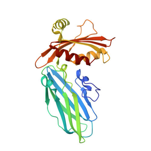
    1QTP, 1QTS

  • PubMed Abstract: 

    AP-2 adaptors regulate clathrin-bud formation at the cell surface by recruiting clathrin trimers to the plasma membrane and by selecting certain membrane proteins for inclusion within the developing clathrin-coat structure. These functions are performed by discrete subunits of the adaptor heterotetramer. The carboxyl-terminal appendage of the AP-2 alpha subunit appears to regulate the translocation of several endocytic accessory proteins to the bud site. We have determined the crystal structure of the alpha appendage at 1.4-A resolution by multiwavelength anomalous diffraction phasing. It is composed of two distinct structural modules, a beta-sandwich domain and a mixed alpha-beta platform domain. Structure-based mutagenesis shows that alterations to the molecular surface of a highly conserved region on the platform domain differentially affect associations of the appendage with amphiphysin, eps15, epsin, and AP180, revealing a common protein-binding interface.


  • Organizational Affiliation

    Department of Internal Medicine, Washington University School of Medicine, 660 South Euclid Avenue, St. Louis, MO 63110, USA.


Macromolecules
Find similar proteins by:  (by identity cutoff)  |  3D Structure
Entity ID: 1
MoleculeChains Sequence LengthOrganismDetailsImage
AP-2 CLATHRIN ADAPTOR ALPHA SUBUNIT (ALPHA-ADAPTIN C)247Mus musculusMutation(s): 0 
UniProt & NIH Common Fund Data Resources
Find proteins for P17427 (Mus musculus)
Explore P17427 
Go to UniProtKB:  P17427
IMPC:  MGI:101920
Entity Groups  
Sequence Clusters30% Identity50% Identity70% Identity90% Identity95% Identity100% Identity
UniProt GroupP17427
Sequence Annotations
Expand
  • Reference Sequence
Experimental Data & Validation

Experimental Data

  • Method: X-RAY DIFFRACTION
  • Resolution: 1.40 Å
  • R-Value Free: 0.209 
  • R-Value Work: 0.178 
  • Space Group: P 1 21 1
Unit Cell:
Length ( Å )Angle ( ˚ )
a = 40.807α = 90
b = 72.749β = 99.69
c = 41.861γ = 90
Software Package:
Software NamePurpose
AMoREphasing
REFMACrefinement
DENZOdata reduction
SCALEPACKdata scaling

Structure Validation

View Full Validation Report


Entry History 

Deposition Data

Revision History  (Full details and data files)

  • Version 1.0: 1999-07-12
    Type: Initial release
  • Version 1.1: 2008-04-27
    Changes: Version format compliance
  • Version 1.2: 2011-07-13
    Changes: Version format compliance
  • Version 1.3: 2024-02-14
    Changes: Data collection, Database references