1QMG

Acetohydroxyacid isomeroreductase complexed with its reaction product dihydroxy-methylvalerate, manganese and ADP-ribose.


Experimental Data Snapshot

  • Method: X-RAY DIFFRACTION
  • Resolution: 1.60 Å
  • R-Value Free: 0.224 
  • R-Value Work: 0.196 
  • R-Value Observed: 0.196 

wwPDB Validation   3D Report Full Report


Ligand Structure Quality Assessment 


This is version 1.3 of the entry. See complete history


Literature

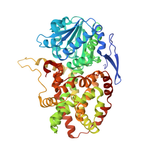
Structure of Spinach Acetohydroxyacid Isomeroreductase Complexed with its Product of Reaction Dihydroxy-Methylvalerate, Manganese and Adp-Ribose

Thomazeau, K.Dumas, R.Halgand, F.Forest, E.Douce, R.Biou, V.

(2000) Acta Crystallogr D Biol Crystallogr 56: 389

  • DOI: https://doi.org/10.1107/s0907444900001694
  • Primary Citation of Related Structures:  
    1QMG

  • PubMed Abstract: 

    Acetohydroxyacid isomeroreductase catalyses a two-step reaction composed of an alkyl migration followed by an NADPH-dependent reduction. Both steps require a divalent cation and the first step has a strong preference for magnesium. Manganese ions are highly unfavourable to the reaction: only 3% residual activity is observed in the presence of this cation. Acetohydroxyacid isomeroreductase has been crystallized with its substrate, 2-aceto-2-hydroxybutyrate (AHB), Mn(2+) and NADPH. The 1.6 A resolution electron-density map showed the reaction product (2,3-dihydroxy-3-methylvalerate, DHMV) and a density corresponding to (phospho)-ADP-ribose instead of the whole NADP(+). This is one of the few structures of an enzyme complexed with its reaction product. The structure of this complex was refined to an R factor of 19.3% and an R(free) of 22.5%. The overall structure of the enzyme is very similar to that of the complex with the reaction-intermediate analogue IpOHA [N-hydroxy-N-isopropyloxamate; Biou et al. (1997), EMBO J. 16, 3405-3415]. However, the active site shows some differences: the nicotinamide is cleaved and the surrounding amino acids have rearranged accordingly. Comparison between the structures corresponding to the reaction intermediate and to the end of the reaction allowed the proposal of a reaction scheme. Taking this result into account, the enzyme was crystallized with Ni(2+) and Zn(2+), for which only 0.02% residual activity were measured; however, the crystals of AHB/Zn/NADPH and of AHB/Ni/NADPH also contain the reaction product. Moreover, mass-spectrometry measurements confirmed the -cleavage of nicotinamide.


  • Organizational Affiliation

    Institut de Biologie Structurale Jean-Pierre Ebel (UMR 5045), CNRS/CEA/Université Joseph Fourier, 41 Rue Jules Horowitz, F-38027 Grenoble CEDEX, France.


Macromolecules
Find similar proteins by:  (by identity cutoff)  |  3D Structure
Entity ID: 1
MoleculeChains Sequence LengthOrganismDetailsImage
ACETOHYDROXY-ACID ISOMEROREDUCTASE
A, B, C, D
524Spinacia oleraceaMutation(s): 0 
EC: 1.1.1.86
UniProt
Find proteins for Q01292 (Spinacia oleracea)
Explore Q01292 
Go to UniProtKB:  Q01292
Entity Groups  
Sequence Clusters30% Identity50% Identity70% Identity90% Identity95% Identity100% Identity
UniProt GroupQ01292
Sequence Annotations
Expand
  • Reference Sequence
Small Molecules
Ligands 4 Unique
IDChains Name / Formula / InChI Key2D Diagram3D Interactions
APX
Query on APX

Download Ideal Coordinates CCD File 
E [auth A],
J [auth B],
N [auth C],
R [auth D]
2'-MONOPHOSPHOADENOSINE-5'-DIPHOSPHORIBOSE
C15 H29 N5 O17 P3
QHNQLFGTVLWISK-MQSGHBOVSA-O
DMV
Query on DMV

Download Ideal Coordinates CCD File 
I [auth A],
M [auth B],
Q [auth C],
U [auth D]
2,3-DIHYDROXY-VALERIANIC ACID
C6 H12 O4
PDGXJDXVGMHUIR-UJURSFKZSA-N
SO4
Query on SO4

Download Ideal Coordinates CCD File 
H [auth A]SULFATE ION
O4 S
QAOWNCQODCNURD-UHFFFAOYSA-L
MN
Query on MN

Download Ideal Coordinates CCD File 
F [auth A]
G [auth A]
K [auth B]
L [auth B]
O [auth C]
F [auth A],
G [auth A],
K [auth B],
L [auth B],
O [auth C],
P [auth C],
S [auth D],
T [auth D]
MANGANESE (II) ION
Mn
WAEMQWOKJMHJLA-UHFFFAOYSA-N
Experimental Data & Validation

Experimental Data

  • Method: X-RAY DIFFRACTION
  • Resolution: 1.60 Å
  • R-Value Free: 0.224 
  • R-Value Work: 0.196 
  • R-Value Observed: 0.196 
  • Space Group: P 1 21 1
Unit Cell:
Length ( Å )Angle ( ˚ )
a = 110.8α = 90
b = 61.2β = 95
c = 161.7γ = 90
Software Package:
Software NamePurpose
X-PLORrefinement
MOSFLMdata reduction
SCALAdata scaling
X-PLORphasing

Structure Validation

View Full Validation Report


Ligand Structure Quality Assessment 


Entry History 

Deposition Data

Revision History  (Full details and data files)

  • Version 1.0: 2000-03-31
    Type: Initial release
  • Version 1.1: 2011-05-08
    Changes: Version format compliance
  • Version 1.2: 2011-07-13
    Changes: Version format compliance
  • Version 1.3: 2023-12-13
    Changes: Data collection, Database references, Derived calculations, Other, Refinement description