1QJC

Phosphopantetheine Adenylyltransferase from Escherichia coli in complex with 4'-phosphopantetheine


Experimental Data Snapshot

  • Method: X-RAY DIFFRACTION
  • Resolution: 1.63 Å
  • R-Value Free: 0.247 
  • R-Value Work: 0.216 
  • R-Value Observed: 0.216 

wwPDB Validation   3D Report Full Report


This is version 1.1 of the entry. See complete history


Literature

The Crystal Structures of Phosphopantetheine Adenylyltransferase with Bound Substrates Reveal the Enzyme'S Catalytic Mechanism

Izard, T.

(2001) J Mol Biol 315: 487

  • DOI: https://doi.org/10.1006/jmbi.2001.5272
  • Primary Citation of Related Structures:  
    1GN8, 1QJC

  • PubMed Abstract: 

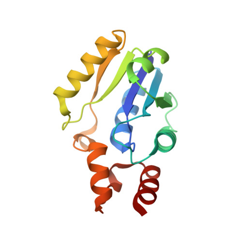
    Phosphopantetheine adenylyltransferase (PPAT) is an essential enzyme in the coenzyme A pathway that catalyzes the reversible transfer of an adenylyl group from ATP to 4'-phosphopantetheine (Ppant) in the presence of magnesium. To investigate the reaction mechanism, the high-resolution crystal structures of the Escherichia coli PPAT have been determined in the presence of either ATP or Ppant. Structural details of the catalytic center revealed specific roles for individual amino acid residues involved in substrate binding and catalysis. The side-chain of His18 stabilizes the expected pentacovalent intermediate, whereas the side-chains of Thr10 and Lys42 orient the nucleophile for an in-line displacement mechanism. The binding site for the manganese ion that interacts with the phosphate groups of the nucleotide has also been identified. Within the PPAT hexamer, one trimer is in its substrate-free state, whereas the other is in a substrate-bound state.


  • Organizational Affiliation

    Department of Structural Biology, St. Jude Children's Research Hospital, 332 North Lauderdale Street, Memphis, TN 38105-2794, USA. Tina.Izard@stjude.org


Macromolecules
Find similar proteins by:  (by identity cutoff)  |  3D Structure
Entity ID: 1
MoleculeChains Sequence LengthOrganismDetailsImage
PHOSPHOPANTETHEINE ADENYLYLTRANSFERASE
A, B
158Escherichia coliMutation(s): 0 
EC: 2.7.7.3
UniProt
Find proteins for P0A6I6 (Escherichia coli (strain K12))
Explore P0A6I6 
Go to UniProtKB:  P0A6I6
Entity Groups  
Sequence Clusters30% Identity50% Identity70% Identity90% Identity95% Identity100% Identity
UniProt GroupP0A6I6
Sequence Annotations
Expand
  • Reference Sequence
Experimental Data & Validation

Experimental Data

  • Method: X-RAY DIFFRACTION
  • Resolution: 1.63 Å
  • R-Value Free: 0.247 
  • R-Value Work: 0.216 
  • R-Value Observed: 0.216 
  • Space Group: I 2 3
Unit Cell:
Length ( Å )Angle ( ˚ )
a = 135.331α = 90
b = 135.331β = 90
c = 135.331γ = 90
Software Package:
Software NamePurpose
CNSrefinement
DENZOdata reduction
SCALEPACKdata scaling

Structure Validation

View Full Validation Report


Entry History 

Deposition Data

  • Released Date: 2001-06-27 
  • Deposition Author(s): Izard, T.

Revision History  (Full details and data files)

  • Version 1.0: 2001-06-27
    Type: Initial release
  • Version 1.1: 2023-12-13
    Changes: Data collection, Database references, Refinement description