1QFK

STRUCTURE OF HUMAN FACTOR VIIA AND ITS IMPLICATIONS FOR THE TRIGGERING OF BLOOD COAGULATION


Experimental Data Snapshot

  • Method: X-RAY DIFFRACTION
  • Resolution: 2.80 Å
  • R-Value Free: 0.267 
  • R-Value Work: 0.215 

wwPDB Validation   3D Report Full Report


Ligand Structure Quality Assessment 


This is version 2.1 of the entry. See complete history


Literature

Structure of human factor VIIa and its implications for the triggering of blood coagulation.

Pike, A.C.Brzozowski, A.M.Roberts, S.M.Olsen, O.H.Persson, E.

(1999) Proc Natl Acad Sci U S A 96: 8925-8930

  • DOI: https://doi.org/10.1073/pnas.96.16.8925
  • Primary Citation of Related Structures:  
    1QFK

  • PubMed Abstract: 

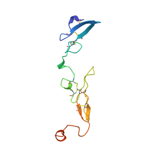
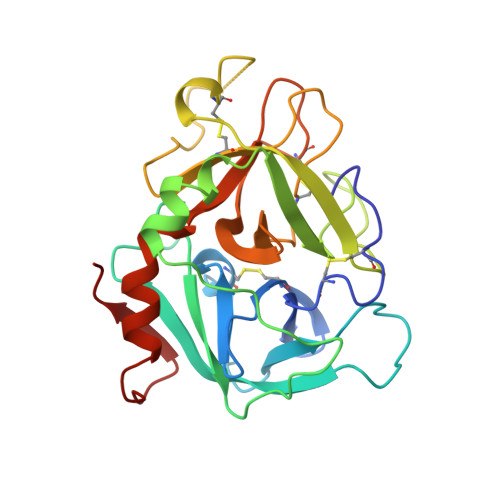
    Factor VIIa (EC 3.4.21.21) is a trypsin-like serine protease that plays a key role in the blood coagulation cascade. On injury, factor VIIa forms a complex with its allosteric regulator, tissue factor, and initiates blood clotting. Although the structure of the binary complex has already been determined [Banner, D. W., D'Arcy, A., Chène, C., Winkler, F. K., Guha, A., Konigsberg, W. H., Nemerson, Y. & Kirchhofer, D. (1996) Nature (London) 380, 41-46], the conformational effects of cofactor binding to factor VIIa are not known in detail because of a lack of structural information on free factor VIIa. Here we report the structure of gamma-carboxyglutamic acid-domainless human coagulation factor VIIa at a resolution of 2.8 A. The molecule adopts an extended conformation within the crystal similar to that previously observed for the full-length protein in complex with tissue factor. Detailed comparison of free and tissue factor-bound factor VIIa reveals several structural differences. The binding mode of the active-site inhibitor D-Phe-Phe-Arg methyl ketone differs in the two structures, suggesting a role for the cofactor in substrate recognition. More importantly, a surface-exposed alpha-helix in the protease domain (residues 307-312), which is located at the cofactor recognition site, is distorted in the free form of factor VIIa. This subtle structural difference sheds light on the mechanism of the dramatic tissue factor-induced enhancement of factor VIIa activity.


  • Organizational Affiliation

    Structural Biology Laboratory, Chemistry Department, University of York, York YO10 5DD, United Kingdom.


Macromolecules
Find similar proteins by:  (by identity cutoff)  |  3D Structure
Entity ID: 1
MoleculeChains Sequence LengthOrganismDetailsImage
COAGULATION FACTOR VIIA LIGHT CHAINA [auth L]104Homo sapiensMutation(s): 0 
EC: 3.4.21.21
UniProt & NIH Common Fund Data Resources
Find proteins for P08709 (Homo sapiens)
Explore P08709 
Go to UniProtKB:  P08709
PHAROS:  P08709
GTEx:  ENSG00000057593 
Entity Groups  
Sequence Clusters30% Identity50% Identity70% Identity90% Identity95% Identity100% Identity
UniProt GroupP08709
Sequence Annotations
Expand
  • Reference Sequence
Find similar proteins by:  (by identity cutoff)  |  3D Structure
Entity ID: 2
MoleculeChains Sequence LengthOrganismDetailsImage
COAGULATION FACTOR VIIA HEAVY CHAINB [auth H]254Homo sapiensMutation(s): 0 
EC: 3.4.21.21
UniProt & NIH Common Fund Data Resources
Find proteins for P08709 (Homo sapiens)
Explore P08709 
Go to UniProtKB:  P08709
PHAROS:  P08709
GTEx:  ENSG00000057593 
Entity Groups  
Sequence Clusters30% Identity50% Identity70% Identity90% Identity95% Identity100% Identity
UniProt GroupP08709
Sequence Annotations
Expand
  • Reference Sequence
Oligosaccharides

Help

Entity ID: 3
MoleculeChains Length2D Diagram Glycosylation3D Interactions
2-acetamido-2-deoxy-beta-D-glucopyranose-(1-4)-[beta-L-fucopyranose-(1-6)]2-acetamido-2-deoxy-beta-D-glucopyranoseC [auth A]3N-Glycosylation
Glycosylation Resources
GlyTouCan:  G28454KX
GlyCosmos:  G28454KX
GlyGen:  G28454KX
Small Molecules
Ligands 4 Unique
IDChains Name / Formula / InChI Key2D Diagram3D Interactions
0Z6
Query on 0Z6

Download Ideal Coordinates CCD File 
F [auth H]D-phenylalanyl-N-[(2S,3S)-6-{[amino(iminio)methyl]amino}-1-chloro-2-hydroxyhexan-3-yl]-L-phenylalaninamide
C25 H36 Cl N6 O3
ZKHBINZTIMXMQW-CLAROIROSA-O
GLC
Query on GLC

Download Ideal Coordinates CCD File 
D [auth L]alpha-D-glucopyranose
C6 H12 O6
WQZGKKKJIJFFOK-DVKNGEFBSA-N
FUC
Query on FUC

Download Ideal Coordinates CCD File 
E [auth L]alpha-L-fucopyranose
C6 H12 O5
SHZGCJCMOBCMKK-SXUWKVJYSA-N
CA
Query on CA

Download Ideal Coordinates CCD File 
G [auth H]CALCIUM ION
Ca
BHPQYMZQTOCNFJ-UHFFFAOYSA-N
Biologically Interesting Molecules (External Reference) 1 Unique
Experimental Data & Validation

Experimental Data

  • Method: X-RAY DIFFRACTION
  • Resolution: 2.80 Å
  • R-Value Free: 0.267 
  • R-Value Work: 0.215 
  • Space Group: P 43 2 2
Unit Cell:
Length ( Å )Angle ( ˚ )
a = 115.3α = 90
b = 115.3β = 90
c = 98γ = 90
Software Package:
Software NamePurpose
DENZOdata reduction
SCALEPACKdata scaling
AMoREphasing
REFMACrefinement

Structure Validation

View Full Validation Report


Ligand Structure Quality Assessment 


Entry History 

Deposition Data

Revision History  (Full details and data files)

  • Version 1.0: 1999-08-01
    Type: Initial release
  • Version 1.1: 2007-10-16
    Changes: Version format compliance
  • Version 1.2: 2011-07-13
    Changes: Atomic model, Database references, Derived calculations, Non-polymer description, Structure summary, Version format compliance
  • Version 1.3: 2012-12-12
    Changes: Other
  • Version 2.0: 2020-07-29
    Type: Remediation
    Reason: Carbohydrate remediation
    Changes: Advisory, Atomic model, Data collection, Derived calculations, Structure summary
  • Version 2.1: 2023-08-16
    Changes: Data collection, Database references, Refinement description, Structure summary