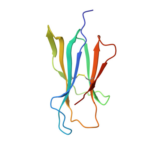
beta2-microglobulin H31Y variant 3D structure highlights the protein natural propensity towards intermolecular aggregation
Rosano, C., Zuccotti, S., Mangione, P., Giorgetti, S., Bellotti, V., Pettirossi, F., Corazza, A., Viglino, P., Esposito, G., Bolognesi, M.(2004) J Mol Biol 335: 1051-1064
- PubMed: 14698299
- DOI: https://doi.org/10.1016/j.jmb.2003.11.040
- Primary Citation of Related Structures:
1PY4 - PubMed Abstract:
beta2-Microglobulin (beta2m) is the non-covalently bound light chain of the human class I major histocompatibility complex (MHC-I). The natural turnover of MHC-I gives rise to the release of beta2m into plasmatic fluids and to its catabolism in the kidney. beta2m dissociation from the heavy chain of the complex is a severe complication in patients receiving prolonged hemodialysis. As a consequence of renal failure, the increasing beta2m concentrations can lead to deposition of the protein as amyloid fibrils. Here we characterize the His31-->Tyr human beta2m mutant, a non-natural form of beta2m that is more stable than the wild-type protein, displaying a ten-fold acceleration of the slow phase of folding. We report the 2.9A resolution crystal structure and the NMR characterization of the mutant beta2m, focussing on selected structural features and on the molecular packing observed in the crystals. Juxtaposition of the four mutant beta2m molecules contained in the crystal asymmetric unit, and specific hydrogen bonds, stabilize a compact protein assembly. Conformational heterogeneity of the four independent molecules, some of their mutual interactions and partial unpairing of the N-terminal beta-strand in one protomer are in keeping with the amyloidogenic properties displayed by the mutant beta2m.
Organizational Affiliation:
Istituto Nazionale Ricerca sul Cancro-IST, X-ray Structural Biology Unit, Largo Rosanno Benzi 10, 16132 Genova, Italy.