1PVS

3-methyladenine Glcosylase II(AlkA) Hypoxanthine complex


Experimental Data Snapshot

  • Method: X-RAY DIFFRACTION
  • Resolution: 2.40 Å
  • R-Value Free: 0.292 
  • R-Value Work: 0.232 

wwPDB Validation   3D Report Full Report


This is version 1.3 of the entry. See complete history


Literature

3-methyladenine-DNA glycosylase II: the crystal structure of an AlkA-hypoxanthine complex suggests the possibility of product inhibition.

Teale, M.Symersky, J.DeLucas, L.

(2002) Bioconjug Chem 13: 403-407

  • DOI: https://doi.org/10.1021/bc015527v
  • Primary Citation of Related Structures:  
    1PVS

  • PubMed Abstract: 

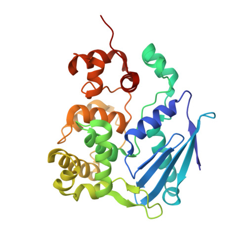
    Escherichia coli (E. coli) protein 3-methyladenine-DNA glycosylase II (AlkA) functions primarily by removing alkylation damage from duplex and single stranded DNA. A crystal structure of AlkA was refined to 2.0 A resolution. This structure in turn was used to refine an AlkA-hypoxanthine (substrate) complex structure to 2.4 A resolution. The complex structure shows hypoxanthine located in AlkA's active site stacked between residues W218 and Y239. The structural analysis of the AlkA and AlkA-hypoxanthine structures indicate that free hypoxanthine binding in the active site may inhibit glycosylase activity.


  • Organizational Affiliation

    Laboratory for Structural Biology, University of Alabama in Huntsville, 35899, USA. tealem@email.uah.edu


Macromolecules
Find similar proteins by:  (by identity cutoff)  |  3D Structure
Entity ID: 1
MoleculeChains Sequence LengthOrganismDetailsImage
DNA-3-methyladenine glycosylase II
A, B
282Escherichia coliMutation(s): 0 
Gene Names: ALKA OR AIDA OR B2068
EC: 3.2.2.21
UniProt
Find proteins for P04395 (Escherichia coli (strain K12))
Explore P04395 
Go to UniProtKB:  P04395
Entity Groups  
Sequence Clusters30% Identity50% Identity70% Identity90% Identity95% Identity100% Identity
UniProt GroupP04395
Sequence Annotations
Expand
  • Reference Sequence
Small Molecules
Ligands 1 Unique
IDChains Name / Formula / InChI Key2D Diagram3D Interactions
7HP
Query on 7HP

Download Ideal Coordinates CCD File 
C [auth A],
D [auth B]
7-HYDROXY-PYRAZOLO[4,3-D]PYRIMIDINE
C5 H4 N4 O
JFZSDNLQDTYVEE-UHFFFAOYSA-N
Experimental Data & Validation

Experimental Data

  • Method: X-RAY DIFFRACTION
  • Resolution: 2.40 Å
  • R-Value Free: 0.292 
  • R-Value Work: 0.232 
  • Space Group: P 1 21 1
Unit Cell:
Length ( Å )Angle ( ˚ )
a = 58.005α = 90
b = 75.979β = 110.03
c = 61.417γ = 90
Software Package:
Software NamePurpose
CNSrefinement
DENZOdata reduction
SCALEPACKdata scaling
X-PLORphasing

Structure Validation

View Full Validation Report


Entry History 

Deposition Data

  • Released Date: 2004-06-08 
  • Deposition Author(s): Teale, M.

Revision History  (Full details and data files)

  • Version 1.0: 2004-06-08
    Type: Initial release
  • Version 1.1: 2008-04-29
    Changes: Version format compliance
  • Version 1.2: 2011-07-13
    Changes: Version format compliance
  • Version 1.3: 2024-02-14
    Changes: Data collection, Database references, Derived calculations