Crystallization and structure determination of bovine profilin at 2.0 A resolution.
Cedergren-Zeppezauer, E.S., Goonesekere, N.C., Rozycki, M.D., Myslik, J.C., Dauter, Z., Lindberg, U., Schutt, C.E.(1994) J Mol Biol 240: 459-475
- PubMed: 8046751
- DOI: https://doi.org/10.1006/jmbi.1994.1461
- Primary Citation of Related Structures:
1PNE - PubMed Abstract:
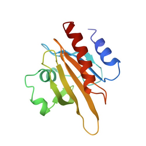
Profilin regulates the behavior of the eukaryotic microfilament system through its interaction with non-filamentous actin. It also binds several ligands, including poly(L-proline) and the membrane phospholipid phosphatidylinositol 4,5-bisphosphate (PtdIns(4,5)P2). Bovine profilin crystals (space group C2; a = 69.15 A, b = 34.59 A, c = 52.49 A; alpha = gamma = 90 degrees, beta = 92.56 degrees) were grown from a mixture of poly(ethylene glycol) 400 and ammonium sulfate. X-ray diffraction data were collected on an imaging plate scanner at the DORIS storage ring (DESY, Hamburg), and were phased by molecular replacement, using a search model derived from the 2.55 A structure of profilin complexed to beta-actin. The refined model of bovine profilin has a crystallographic R-factor of 16.5% in the resolution range 6.0 to 2.0 A and includes 128 water molecules, several of which form hydrogen bonds to stabilize unconventional turns. The structure of free bovine profilin is similar to that of bovine profilin complexed to beta-actin, and C alpha atoms from the two structures superimpose with an r.m.s. deviation of 1.25 A. This value is reduced to 0.51 A by omitting Ala1 and the N-terminal acetyl group, which lie at a profilin-actin interface in crystals of the complex. These residues display a strained conformation in crystalline profilin-actin but may allow the formation of a hydrogen bond between the N-acetyl carbonyl group of profilin and the phenol hydroxyl group of Tyr188 in actin. Several other actin-binding residues of profilin show different side-chain rotomer conformations in the two structures. The polypeptide fold of bovine profilin is generally similar to those observed by NMR for profilin from other sources, although the N terminus of Acanthamoeba profilin isoform I lies in a distorted helix and the C-terminal helix is less tilted with respect to the strands in the central beta-pleated sheet than is observed in bovine profilin. The majority of the aromatic residues in profilin are exposed to solvent and lie in either of two hydrophobic patches, neither of which takes part in an interface with actin. One of these patches is required for binding poly(L-proline) and contains an aromatic cluster comprising the highly conserved residues Trp3, Tyr6, Trp31 and Tyr139. In forming this cluster, Trp31 adopts a sterically strained rotamer conformation.(ABSTRACT TRUNCATED AT 400 WORDS)
Organizational Affiliation:
Department of Zoological Cell Biology, Arrhenius Laboratories for Natural Sciences, Stockholm University, Sweden.