1PEK

STRUCTURE OF THE COMPLEX OF PROTEINASE K WITH A SUBSTRATE-ANALOGUE HEXA-PEPTIDE INHIBITOR AT 2.2 ANGSTROMS RESOLUTION


Experimental Data Snapshot

  • Method: X-RAY DIFFRACTION
  • Resolution: 2.20 Å
  • R-Value Observed: 0.165 

wwPDB Validation   3D Report Full Report


This is version 1.3 of the entry. See complete history


Literature

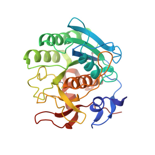
Structure of the complex of proteinase K with a substrate analogue hexapeptide inhibitor at 2.2-A resolution.

Betzel, C.Singh, T.P.Visanji, M.Peters, K.Fittkau, S.Saenger, W.Wilson, K.S.

(1993) J Biol Chem 268: 15854-15858

  • Primary Citation of Related Structures:  
    1PEK

  • PubMed Abstract: 

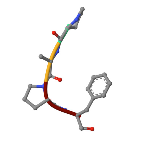
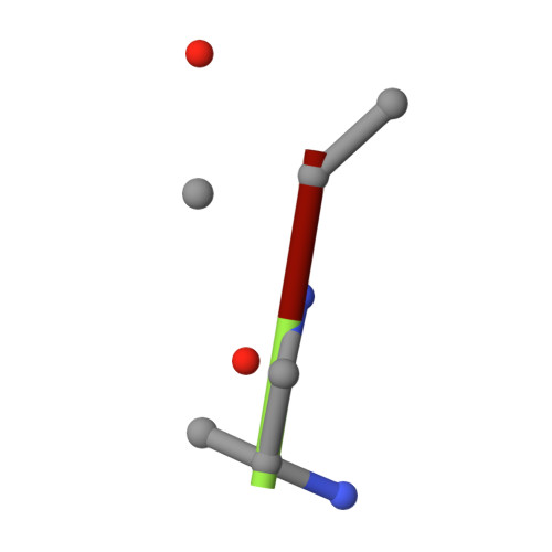
    The crystal structure of a transition state/product complex formed by the interaction between proteinase K and the substrate analogue N-Ac-L-Pro-L-Ala-L-Pro-L-Phe-D-Ala-L-Ala-NH2 has been determined at a resolution of 2.2 A and refined to an R-factor of 0.165 for 12,725 reflections. The inhibitor forms a stable complex through a series of hydrogen bonds with protein atoms and water molecules. The inhibitor is hydrolyzed between Phe 4I and D-Ala5I (I indicates inhibitor). The two fragments are separated by a distance of 3.07 A between the carbonyl carbon and the main chain nitrogen. Both fragments remain bound to the protein. The N-terminal fragment occupies subsites S5 to S1, whereas the C-terminal part is bound in S1' and S2', the first time that electron density for a substrate analogue has been observed in the P1' and P2' sites of a subtilisin-like enzyme. The flexible segments of the substrate recognition sites Gly100-Tyr104 and Ser132-Gly136 move appreciably to accommodate the inhibitor. Biochemical results indicate an inhibition by this specifically designed peptide of 95%.


  • Organizational Affiliation

    European Molecular Biology Laboratory, Hamburg, Germany.


Macromolecules
Find similar proteins by:  (by identity cutoff)  |  3D Structure
Entity ID: 1
MoleculeChains Sequence LengthOrganismDetailsImage
PROTEINASE KA [auth E]279Parengyodontium albumMutation(s): 0 
EC: 3.4.21.64
UniProt
Find proteins for P06873 (Parengyodontium album)
Explore P06873 
Go to UniProtKB:  P06873
Entity Groups  
Sequence Clusters30% Identity50% Identity70% Identity90% Identity95% Identity100% Identity
UniProt GroupP06873
Sequence Annotations
Expand
  • Reference Sequence

Find similar proteins by:  Sequence   |   3D Structure  

Entity ID: 2
MoleculeChains Sequence LengthOrganismDetailsImage
PEPTIDE PRO-ALA-PRO-PHEB [auth C]4N/AMutation(s): 0 
Sequence Annotations
Expand
  • Reference Sequence

Find similar proteins by:  Sequence   |   3D Structure  

Entity ID: 3
MoleculeChains Sequence LengthOrganismDetailsImage
D-DAL-ALA-NH2C [auth D]3N/AMutation(s): 0 
Sequence Annotations
Expand
  • Reference Sequence
Experimental Data & Validation

Experimental Data

  • Method: X-RAY DIFFRACTION
  • Resolution: 2.20 Å
  • R-Value Observed: 0.165 
  • Space Group: P 43 21 2
Unit Cell:
Length ( Å )Angle ( ˚ )
a = 68.28α = 90
b = 68.28β = 90
c = 107.87γ = 90
Software Package:
Software NamePurpose
PROLSQrefinement

Structure Validation

View Full Validation Report


Entry History 

Deposition Data

Revision History  (Full details and data files)

  • Version 1.0: 1994-01-31
    Type: Initial release
  • Version 1.1: 2008-03-03
    Changes: Version format compliance
  • Version 1.2: 2011-07-13
    Changes: Atomic model, Database references, Derived calculations, Non-polymer description, Structure summary, Version format compliance
  • Version 1.3: 2017-11-29
    Changes: Advisory, Derived calculations, Other