


Crystal structure of E.coli chloramphenicol acetyltransferase type I at 2.5 Angstrom resolution
Roidis, A., Kokkinidis, M.To be published.
Experimental Data Snapshot
wwPDB Validation 3D Report Full Report

Entity ID: 1 | |||||
|---|---|---|---|---|---|
| Molecule | Chains | Sequence Length | Organism | Details | Image |
| Chloramphenicol acetyltransferase | 219 | Escherichia coli | Mutation(s): 0 Gene Names: CAT OR HCM1.206 EC: 2.3.1.28 | 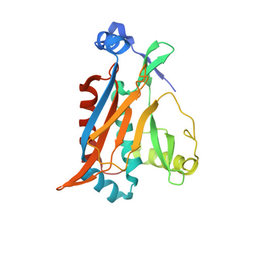 | |
UniProt | |||||
Find proteins for P62577 (Escherichia coli) Explore P62577 Go to UniProtKB: P62577 | |||||
Entity Groups | |||||
| Sequence Clusters | 30% Identity50% Identity70% Identity90% Identity95% Identity100% Identity | ||||
| UniProt Group | P62577 | ||||
Sequence AnnotationsExpand | |||||
| |||||
| Length ( Å ) | Angle ( ˚ ) |
|---|---|
| a = 115.739 | α = 90 |
| b = 129.702 | β = 108.38 |
| c = 117.984 | γ = 90 |
| Software Name | Purpose |
|---|---|
| REFMAC | refinement |
| MAR345 | data collection |
| SCALEPACK | data scaling |
| AMoRE | phasing |
RCSB PDB (citation) is hosted by
RCSB PDB is a member of the

