1PBQ

CRYSTAL STRUCTURE OF THE NR1 LIGAND BINDING CORE IN COMPLEX WITH 5,7-DICHLOROKYNURENIC ACID (DCKA) AT 1.90 ANGSTROMS RESOLUTION


Experimental Data Snapshot

  • Method: X-RAY DIFFRACTION
  • Resolution: 1.90 Å
  • R-Value Free: 0.270 
  • R-Value Work: 0.231 
  • R-Value Observed: 0.231 

wwPDB Validation   3D Report Full Report


This is version 1.4 of the entry. See complete history


Literature

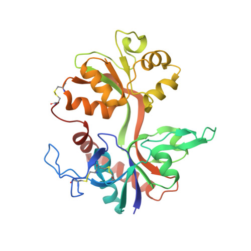
Mechanisms of activation, inhibition and specificity: crystal structures of the NMDA receptor NR1 ligand-binding core

Furukawa, H.Gouaux, E.

(2003) EMBO J 22: 2873-2885

  • DOI: https://doi.org/10.1093/emboj/cdg303
  • Primary Citation of Related Structures:  
    1PB7, 1PB8, 1PB9, 1PBQ

  • PubMed Abstract: 

    Excitatory neurotransmission mediated by the N-methyl-D-aspartate subtype of ionotropic glutamate receptors is fundamental to the development and function of the mammalian central nervous system. NMDA receptors require both glycine and glutamate for activation with NR1 and NR2 forming glycine and glutamate sites, respectively. Mechanisms to describe agonist and antagonist binding, and activation and desensitization of NMDA receptors have been hampered by the lack of high-resolution structures. Here, we describe the cocrystal structures of the NR1 S1S2 ligand-binding core with the agonists glycine and D-serine (DS), the partial agonist D-cycloserine (DCS) and the antagonist 5,7-dichlorokynurenic acid (DCKA). The cleft of the S1S2 'clamshell' is open in the presence of the antagonist DCKA and closed in the glycine, DS and DCS complexes. In addition, the NR1 S1S2 structure reveals the fold and interactions of loop 1, a cysteine-rich region implicated in intersubunit allostery.


  • Organizational Affiliation

    Department of Biochemistry and Molecular Biophysics, Columbia University, 650 West 168th Street, New York, NY 10032, USA.


Macromolecules
Find similar proteins by:  (by identity cutoff)  |  3D Structure
Entity ID: 1
MoleculeChains Sequence LengthOrganismDetailsImage
N-methyl-D-aspartate Receptor Subunit 1
A, B
292Rattus norvegicusMutation(s): 0 
Gene Names: GRIN1 OR NMDAR1
UniProt
Find proteins for P35439 (Rattus norvegicus)
Explore P35439 
Go to UniProtKB:  P35439
Entity Groups  
Sequence Clusters30% Identity50% Identity70% Identity90% Identity95% Identity100% Identity
UniProt GroupP35439
Sequence Annotations
Expand
  • Reference Sequence
Small Molecules
Ligands 1 Unique
IDChains Name / Formula / InChI Key2D Diagram3D Interactions
DK1
Query on DK1

Download Ideal Coordinates CCD File 
C [auth A],
D [auth B]
5,7-DICHLORO-4-HYDROXYQUINOLINE-2-CARBOXYLIC ACID
C10 H5 Cl2 N O3
BGKFPRIGXAVYNX-UHFFFAOYSA-N
Binding Affinity Annotations 
IDSourceBinding Affinity
DK1 PDBBind:  1PBQ Ki: 540 (nM) from 1 assay(s)
BindingDB:  1PBQ Ki: min: 40, max: 90 (nM) from 2 assay(s)
IC50: min: 90, max: 860 (nM) from 3 assay(s)
EC50: 330 (nM) from 1 assay(s)
Binding MOAD:  1PBQ Ki: 540 (nM) from 1 assay(s)
Experimental Data & Validation

Experimental Data

  • Method: X-RAY DIFFRACTION
  • Resolution: 1.90 Å
  • R-Value Free: 0.270 
  • R-Value Work: 0.231 
  • R-Value Observed: 0.231 
  • Space Group: C 1 2 1
Unit Cell:
Length ( Å )Angle ( ˚ )
a = 85.7α = 90
b = 65.3β = 98.1
c = 124.3γ = 90
Software Package:
Software NamePurpose
CNSrefinement
DENZOdata reduction
SCALEPACKdata scaling
AMoREphasing

Structure Validation

View Full Validation Report


Entry History 

Deposition Data

Revision History  (Full details and data files)

  • Version 1.0: 2003-07-29
    Type: Initial release
  • Version 1.1: 2008-04-29
    Changes: Version format compliance
  • Version 1.2: 2011-07-13
    Changes: Version format compliance
  • Version 1.3: 2017-08-16
    Changes: Source and taxonomy
  • Version 1.4: 2020-07-15
    Changes: Advisory, Database references, Derived calculations, Structure summary