1P91

Crystal Structure of RlmA(I) enzyme: 23S rRNA n1-G745 methyltransferase (NORTHEAST STRUCTURAL GENOMICS CONSORTIUM TARGET ER19)


Experimental Data Snapshot

  • Method: X-RAY DIFFRACTION
  • Resolution: 2.80 Å
  • R-Value Free: 0.296 
  • R-Value Work: 0.248 
  • R-Value Observed: 0.248 

wwPDB Validation   3D Report Full Report


Ligand Structure Quality Assessment 


This is version 1.3 of the entry. See complete history


Literature

Crystal structure of RlmAI: implications for understanding the 23S rRNA G745/G748-methylation at the macrolide antibiotic-binding site.

Das, K.Acton, T.Chiang, Y.Shih, L.Arnold, E.Montelione, G.T.

(2004) Proc Natl Acad Sci U S A 101: 4041-4046

  • DOI: https://doi.org/10.1073/pnas.0400189101
  • Primary Citation of Related Structures:  
    1P91

  • PubMed Abstract: 

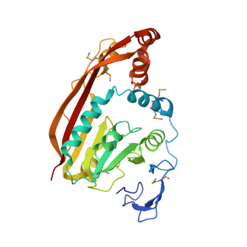
    The RlmA class of enzymes (RlmA(I) and RlmA(II)) catalyzes N1-methylation of a guanine base (G745 in Gram-negative and G748 in Gram-positive bacteria) of hairpin 35 of 23S rRNA. We have determined the crystal structure of Escherichia coli RlmA(I) at 2.8-A resolution, providing 3D structure information for the RlmA class of RNA methyltransferases. The dimeric protein structure exhibits features that provide new insights into its molecular function. Each RlmA(I) molecule has a Zn-binding domain, responsible for specific recognition and binding of its rRNA substrate, and a methyltransferase domain. The asymmetric RlmA(I) dimer observed in the crystal structure has a well defined W-shaped RNA-binding cleft. Two S-adenosyl-l-methionine substrate molecules are located at the two valleys of the W-shaped RNA-binding cleft. The unique shape of the RNA-binding cleft, different from that of known RNA-binding proteins, is highly specific and structurally complements the 3D structure of hairpin 35 of bacterial 23S rRNA. Apart from the hairpin 35, parts of hairpins 33 and 34 also interact with the RlmA(I) dimer.


  • Organizational Affiliation

    Center for Advanced Biotechnology and Medicine, Rutgers University, 679 Hoes Lane, Piscataway, NJ 08854, USA. kalyan@cabm.rutgers.edu


Macromolecules
Find similar proteins by:  (by identity cutoff)  |  3D Structure
Entity ID: 1
MoleculeChains Sequence LengthOrganismDetailsImage
Ribosomal RNA large subunit methyltransferase A
A, B
269Escherichia coliMutation(s): 7 
Gene Names: RRMA OR B1822
EC: 2.1.1.51
UniProt
Find proteins for P36999 (Escherichia coli (strain K12))
Explore P36999 
Go to UniProtKB:  P36999
Entity Groups  
Sequence Clusters30% Identity50% Identity70% Identity90% Identity95% Identity100% Identity
UniProt GroupP36999
Sequence Annotations
Expand
  • Reference Sequence
Small Molecules
Ligands 3 Unique
IDChains Name / Formula / InChI Key2D Diagram3D Interactions
SAM
Query on SAM

Download Ideal Coordinates CCD File 
E [auth A],
K [auth B]
S-ADENOSYLMETHIONINE
C15 H22 N6 O5 S
MEFKEPWMEQBLKI-FCKMPRQPSA-N
SO4
Query on SO4

Download Ideal Coordinates CCD File 
D [auth A],
G [auth B],
H [auth B],
I [auth B],
J [auth B]
SULFATE ION
O4 S
QAOWNCQODCNURD-UHFFFAOYSA-L
ZN
Query on ZN

Download Ideal Coordinates CCD File 
C [auth A],
F [auth B]
ZINC ION
Zn
PTFCDOFLOPIGGS-UHFFFAOYSA-N
Modified Residues  1 Unique
IDChains TypeFormula2D DiagramParent
MSE
Query on MSE
A, B
L-PEPTIDE LINKINGC5 H11 N O2 SeMET
Experimental Data & Validation

Experimental Data

  • Method: X-RAY DIFFRACTION
  • Resolution: 2.80 Å
  • R-Value Free: 0.296 
  • R-Value Work: 0.248 
  • R-Value Observed: 0.248 
  • Space Group: I 2 2 2
Unit Cell:
Length ( Å )Angle ( ˚ )
a = 107.187α = 90
b = 122.279β = 90
c = 143.138γ = 90
Software Package:
Software NamePurpose
CNSrefinement
DENZOdata reduction
SCALEPACKdata scaling
SnBphasing
SOLVEphasing

Structure Validation

View Full Validation Report


Ligand Structure Quality Assessment 


Entry History 

Deposition Data

Revision History  (Full details and data files)

  • Version 1.0: 2004-03-30
    Type: Initial release
  • Version 1.1: 2008-04-29
    Changes: Version format compliance
  • Version 1.2: 2011-07-13
    Changes: Version format compliance
  • Version 1.3: 2017-10-11
    Changes: Refinement description