1ORE

Human Adenine Phosphoribosyltransferase


Experimental Data Snapshot

  • Method: X-RAY DIFFRACTION
  • Resolution: 2.10 Å
  • R-Value Free: 0.176 
  • R-Value Work: 0.133 
  • R-Value Observed: 0.135 

wwPDB Validation   3D Report Full Report


Ligand Structure Quality Assessment 


This is version 1.4 of the entry. See complete history


Literature

Three-dimensional structure of human adenine phosphoribosyltransferase and its relation to DHA-urolithiasis.

Silva, M.Silva, C.H.T.P.Iulek, J.Thiemann, O.H.

(2004) Biochemistry 43: 7663-7671

  • DOI: https://doi.org/10.1021/bi0360758
  • Primary Citation of Related Structures:  
    1ORE

  • PubMed Abstract: 

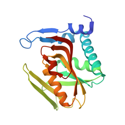
    In mammals, adenine phosphoribosyltransferase (APRT, EC 2.4.2.7) is present in all tissues and provides the only known mechanism for the metabolic salvage of adenine resulting from the polyamine biosynthesis pathway or from dietary sources. In humans, APRT deficiency results in serious kidney illness such as nephrolithiasis, interstitial nephritis, and chronic renal failure as a result of 2,8-dihydroxyadenine (DHA) precipitation in the renal interstitium. To address the molecular basis of DHA-urolithiasis, the recombinant human APRT was crystallized in complex with adenosine 5'-monophosphate (AMP). Refinement of X-ray diffraction data extended to 2.1 A resolution led to a final crystallographic R(factor) of 13.3% and an R(free) of 17.6%. This structure is composed of nine beta-strands and six alpha-helices, and the active site pocket opens slightly to accommodate the AMP product. The core of APRT is similar to that of other phosphoribosyltransferases (PRTases), although the adenine-binding domain is quite different. Structural comparisons between the human APRT and other "type I" PRTases of known structure revealed several important features of the biochemistry of PRTases. We propose that the residues located at positions corresponding to Leu159 and Ala131 in hAPRT are responsible for the base specificities of type I PRTases. The comparative analysis shown here also provides structural information for the mechanism by which mutations in the human APRT lead to DHA-urolithiasis.


  • Organizational Affiliation

    Laboratory of Protein Crystallography and Structural Biology, Physics Institute of São Carlos, University of São Paulo, Av. Trabalhador Sãocarlense 400, P.O. Box 369, 13566-590 São Carlos, SP, Brazil.


Macromolecules
Find similar proteins by:  (by identity cutoff)  |  3D Structure
Entity ID: 1
MoleculeChains Sequence LengthOrganismDetailsImage
Adenine phosphoribosyltransferase180Homo sapiensMutation(s): 0 
Gene Names: APRT
EC: 2.4.2.7
UniProt & NIH Common Fund Data Resources
Find proteins for P07741 (Homo sapiens)
Explore P07741 
Go to UniProtKB:  P07741
PHAROS:  P07741
GTEx:  ENSG00000198931 
Entity Groups  
Sequence Clusters30% Identity50% Identity70% Identity90% Identity95% Identity100% Identity
UniProt GroupP07741
Sequence Annotations
Expand
  • Reference Sequence
Small Molecules
Ligands 2 Unique
IDChains Name / Formula / InChI Key2D Diagram3D Interactions
AMP
Query on AMP

Download Ideal Coordinates CCD File 
C [auth A]ADENOSINE MONOPHOSPHATE
C10 H14 N5 O7 P
UDMBCSSLTHHNCD-KQYNXXCUSA-N
CL
Query on CL

Download Ideal Coordinates CCD File 
B [auth A]CHLORIDE ION
Cl
VEXZGXHMUGYJMC-UHFFFAOYSA-M
Experimental Data & Validation

Experimental Data

  • Method: X-RAY DIFFRACTION
  • Resolution: 2.10 Å
  • R-Value Free: 0.176 
  • R-Value Work: 0.133 
  • R-Value Observed: 0.135 
  • Space Group: P 43 21 2
Unit Cell:
Length ( Å )Angle ( ˚ )
a = 58.309α = 90
b = 58.309β = 90
c = 109.535γ = 90
Software Package:
Software NamePurpose
REFMACrefinement
MAR345data collection
CCP4data scaling
AMoREphasing

Structure Validation

View Full Validation Report


Ligand Structure Quality Assessment 


Entry History 

Deposition Data

Revision History  (Full details and data files)

  • Version 1.0: 2004-03-30
    Type: Initial release
  • Version 1.1: 2008-04-29
    Changes: Version format compliance
  • Version 1.2: 2011-07-13
    Changes: Advisory, Derived calculations, Version format compliance
  • Version 1.3: 2017-10-11
    Changes: Refinement description
  • Version 1.4: 2024-02-14
    Changes: Data collection, Database references, Derived calculations