1OPG

OPG2 FAB FRAGMENT


Experimental Data Snapshot

  • Method: X-RAY DIFFRACTION
  • Resolution: 2.00 Å
  • R-Value Observed: 0.160 

wwPDB Validation   3D Report Full Report


This is version 1.2 of the entry. See complete history


Literature

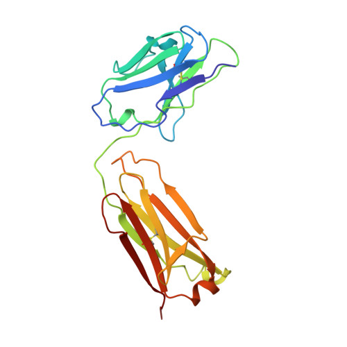
Crystal structure of the OPG2 Fab. An antireceptor antibody that mimics an RGD cell adhesion site.

Kodandapani, R.Veerapandian, B.Kunicki, T.J.Ely, K.R.

(1995) J Biol Chem 270: 2268-2273

  • DOI: https://doi.org/10.1074/jbc.270.5.2268
  • Primary Citation of Related Structures:  
    1OPG

  • PubMed Abstract: 

    Cell surface receptors called integrins mediate diverse cell adhesion phenomena through recognition of the sequence arginine-glycine-aspartic acid (RGD) present in proteins such as fibronectin and fibrinogen. Platelet aggregation in hemostasis is mediated by the binding of fibrinogen to the gpIIb/IIIa integrin. The OPG2 antibody binds the gpIIb/IIIa receptor and acts as a ligand mimic due to the presence of an arginine-tyrosine-aspartic acid (RYD) sequence in the CDR3 loop of the heavy chain. The RYD loop and side chains are ordered in the 2.0-A resolution crystal structure of the Fab fragment from this antireceptor antibody. Moreover, the RYD loop assumes two clearly defined conformations that may correspond to the orientations of the loop in the free state or bound to integrin. This molecule will serve as a tool for understanding protein-integrin recognition in platelet aggregation and other RGD-mediated cell adhesion interactions.


  • Organizational Affiliation

    Cancer Research Center, La Jolla Cancer Research Foundation, California 92037.


Macromolecules
Find similar proteins by:  (by identity cutoff)  |  3D Structure
Entity ID: 1
MoleculeChains Sequence LengthOrganismDetailsImage
OPG2 FAB (LIGHT CHAIN)A [auth L]214Homo sapiensMutation(s): 0 
Entity Groups  
Sequence Clusters30% Identity50% Identity70% Identity90% Identity95% Identity100% Identity
Sequence Annotations
Expand
  • Reference Sequence
Find similar proteins by:  (by identity cutoff)  |  3D Structure
Entity ID: 2
MoleculeChains Sequence LengthOrganismDetailsImage
OPG2 FAB (HEAVY CHAIN)B [auth H]227Homo sapiensMutation(s): 0 
UniProt
Find proteins for P01868 (Mus musculus)
Explore P01868 
Go to UniProtKB:  P01868
Entity Groups  
Sequence Clusters30% Identity50% Identity70% Identity90% Identity95% Identity100% Identity
UniProt GroupP01868
Sequence Annotations
Expand
  • Reference Sequence
Experimental Data & Validation

Experimental Data

  • Method: X-RAY DIFFRACTION
  • Resolution: 2.00 Å
  • R-Value Observed: 0.160 
  • Space Group: P 21 21 2
Unit Cell:
Length ( Å )Angle ( ˚ )
a = 93.1α = 90
b = 83.8β = 90
c = 53.7γ = 90
Software Package:
Software NamePurpose
HOWARDdata collection
A.J.data collection
NILSENdata collection
C.data collection
PROLSQrefinement
XENGENdata reduction
NIELSENdata reduction
XUONG)data reduction

Structure Validation

View Full Validation Report


Entry History 

Deposition Data

Revision History  (Full details and data files)

  • Version 1.0: 1995-07-31
    Type: Initial release
  • Version 1.1: 2008-03-24
    Changes: Version format compliance
  • Version 1.2: 2011-07-13
    Changes: Version format compliance