1OP8

Crystal Structure of Human Granzyme A


Experimental Data Snapshot

  • Method: X-RAY DIFFRACTION
  • Resolution: 2.50 Å
  • R-Value Free: 0.284 
  • R-Value Work: 0.218 

wwPDB Validation   3D Report Full Report


This is version 1.2 of the entry. See complete history


Literature

Crystal structure of the apoptosis-inducing human granzyme A dimer

Hink-Schauer, C.Estebanez-Perpina, E.Kurschus, F.Bode, W.Jenne, D.

(2003) Nat Struct Biol 10: 535-540

  • DOI: https://doi.org/10.1038/nsb945
  • Primary Citation of Related Structures:  
    1OP8

  • PubMed Abstract: 

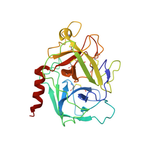
    Granzyme A (GzmA) belongs to a family of trypsin-like serine proteases localized in cytoplasmic granules of activated lymphocytes and natural killer (NK) cells. In contrast to the related granzyme B (GzmB), GzmA forms a stable disulfide-linked homodimer and triggers target-cell death in a caspase-independent way. Limited proteolysis of a high-molecular-mass complex containing SET (also named putative HLA-associated protein II or PHAPII), PHAPI (pp32, leucine-rich acidic nuclear protein) and HMG2 by GzmA liberates NM23-H1, a Mg2+-dependent DNase that causes single-stranded breaks in nuclear DNA. By analyzing the dimeric GzmA structure at a resolution of 2.5 A, we determined the substrate-binding constraints and selective advantages of the two domains arranged as a unique functional tandem. The active sites of the two subunits point in opposite directions and the nearby noncatalytic surfaces can function as exosites, presenting substrates to the active site region of the adjacent partner in a manner analogous to staphylokinase or streptokinase, which present plasminogen to the cofactor-plasmin and cofactor-plasminogen complexes.


  • Organizational Affiliation

    Department of Neuroimmunology, Max Planck Institute of Neurobiology, Am Klopferspitz 18a, D-82152 Planegg-Martinsried, Germany.


Macromolecules
Find similar proteins by:  (by identity cutoff)  |  3D Structure
Entity ID: 1
MoleculeChains Sequence LengthOrganismDetailsImage
Granzyme A
A, B, C, D, E
A, B, C, D, E, F
234Homo sapiensMutation(s): 0 
EC: 3.4.21.78
UniProt & NIH Common Fund Data Resources
Find proteins for P12544 (Homo sapiens)
Explore P12544 
Go to UniProtKB:  P12544
PHAROS:  P12544
GTEx:  ENSG00000145649 
Entity Groups  
Sequence Clusters30% Identity50% Identity70% Identity90% Identity95% Identity100% Identity
UniProt GroupP12544
Sequence Annotations
Expand
  • Reference Sequence
Small Molecules
Ligands 1 Unique
IDChains Name / Formula / InChI Key2D Diagram3D Interactions
SO4
Query on SO4

Download Ideal Coordinates CCD File 
G [auth A]
H [auth A]
I [auth B]
J [auth B]
K [auth B]
G [auth A],
H [auth A],
I [auth B],
J [auth B],
K [auth B],
L [auth C],
M [auth C],
N [auth D],
O [auth D],
P [auth E],
Q [auth F],
R [auth F]
SULFATE ION
O4 S
QAOWNCQODCNURD-UHFFFAOYSA-L
Experimental Data & Validation

Experimental Data

  • Method: X-RAY DIFFRACTION
  • Resolution: 2.50 Å
  • R-Value Free: 0.284 
  • R-Value Work: 0.218 
  • Space Group: P 1
Unit Cell:
Length ( Å )Angle ( ˚ )
a = 49.48α = 117.12
b = 94.55β = 100.25
c = 94.87γ = 100.12
Software Package:
Software NamePurpose
DENZOdata reduction
SCALAdata scaling
AMoREphasing
CNSrefinement
CCP4data scaling

Structure Validation

View Full Validation Report


Entry History 

Deposition Data

Revision History  (Full details and data files)

  • Version 1.0: 2003-07-01
    Type: Initial release
  • Version 1.1: 2008-04-29
    Changes: Version format compliance
  • Version 1.2: 2011-07-13
    Changes: Version format compliance