1OMR

non-myristoylated wild-type bovine recoverin with calcium bound to EF-hand 3


Experimental Data Snapshot

  • Method: X-RAY DIFFRACTION
  • Resolution: 1.50 Å
  • R-Value Free: 0.250 
  • R-Value Work: 0.249 
  • R-Value Observed: 0.249 

wwPDB Validation   3D Report Full Report


This is version 1.3 of the entry. See complete history


Literature

Impact of N-terminal myristoylation on the Ca2+-dependent conformational transition in recoverin

Weiergraber, O.H.Senin, I.I.Philippov, P.P.Granzin, J.Koch, K.-W.

(2003) J Biol Chem 278: 22972-22979

  • DOI: https://doi.org/10.1074/jbc.M300447200
  • Primary Citation of Related Structures:  
    1OMR, 1OMV

  • PubMed Abstract: 

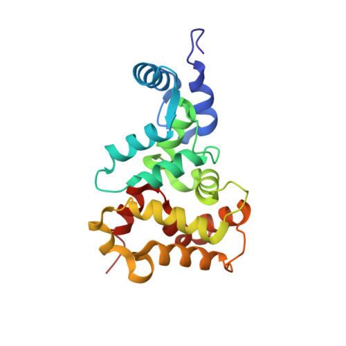
    Recoverin is a Ca2+-regulated signal transduction modulator found in vertebrate retina that has been shown to undergo dramatic conformational changes upon Ca2+ binding to its two functional EF-hand motifs. To elucidate the differential impact of the N-terminal myristoylation as well as occupation of the two Ca2+ binding sites on recoverin structure and function, we have investigated a non-myristoylated E85Q mutant exhibiting virtually no Ca2+ binding to EF-2. Crystal structures of the mutant protein as well as the non-myristoylated wild-type have been determined. Although the non-myristoylated E85Q mutant does not display any functional activity, its three-dimensional structure in the presence of Ca2+ resembles the myristoylated wild-type with two Ca2+ but is quite dissimilar from the myristoylated E85Q mutant. We conclude that the N-terminal myristoyl modification significantly stabilizes the conformation of the Ca2+-free protein (i.e. the T conformation) during the stepwise transition toward the fully Ca2+-occupied state. On the basis of these observations, a refined model for the role of the myristoyl group as an intrinsic allosteric modulator is proposed.


  • Organizational Affiliation

    Institut für Biologische Informationsverarbeitung IBI-2 (Biologische Strukturforschung), Jülich, Germany.


Macromolecules
Find similar proteins by:  (by identity cutoff)  |  3D Structure
Entity ID: 1
MoleculeChains Sequence LengthOrganismDetailsImage
recoverin201Bos taurusMutation(s): 0 
Gene Names: RCV1
UniProt
Find proteins for P21457 (Bos taurus)
Explore P21457 
Go to UniProtKB:  P21457
Entity Groups  
Sequence Clusters30% Identity50% Identity70% Identity90% Identity95% Identity100% Identity
UniProt GroupP21457
Sequence Annotations
Expand
  • Reference Sequence
Small Molecules
Ligands 1 Unique
IDChains Name / Formula / InChI Key2D Diagram3D Interactions
CA
Query on CA

Download Ideal Coordinates CCD File 
B [auth A]CALCIUM ION
Ca
BHPQYMZQTOCNFJ-UHFFFAOYSA-N
Experimental Data & Validation

Experimental Data

  • Method: X-RAY DIFFRACTION
  • Resolution: 1.50 Å
  • R-Value Free: 0.250 
  • R-Value Work: 0.249 
  • R-Value Observed: 0.249 
  • Space Group: I 4
Unit Cell:
Length ( Å )Angle ( ˚ )
a = 84.051α = 90
b = 84.051β = 90
c = 59.179γ = 90
Software Package:
Software NamePurpose
MOSFLMdata reduction
SCALAdata scaling
CNSrefinement
CCP4data scaling
CNSphasing

Structure Validation

View Full Validation Report


Entry History 

Deposition Data

Revision History  (Full details and data files)

  • Version 1.0: 2003-11-25
    Type: Initial release
  • Version 1.1: 2008-04-29
    Changes: Version format compliance
  • Version 1.2: 2011-07-13
    Changes: Derived calculations, Version format compliance
  • Version 1.3: 2023-10-25
    Changes: Data collection, Database references, Derived calculations, Refinement description