1NWD

Solution Structure of Ca2+/Calmodulin bound to the C-terminal Domain of Petunia Glutamate Decarboxylase


Experimental Data Snapshot

  • Method: SOLUTION NMR
  • Conformers Calculated: 200 
  • Conformers Submitted: 20 
  • Selection Criteria: structures with the lowest energy 

wwPDB Validation   3D Report Full Report


This is version 1.3 of the entry. See complete history


Literature

Structural Basis for Simultaneous Binding of Two Carboxy-terminal Peptides of Plant Glutamate Decarboxylase to Calmodulin

Yap, K.L.Yuan, T.Mal, T.K.Vogel, H.J.Ikura, M.

(2003) J Mol Biol 328: 193-204

  • DOI: https://doi.org/10.1016/s0022-2836(03)00271-7
  • Primary Citation of Related Structures:  
    1NWD

  • PubMed Abstract: 

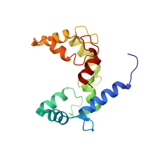
    Activation of glutamate decarboxylase (GAD) by calcium-bound calmodulin (CaM) is required for normal plant growth through regulation of gamma-aminobutyrate and glutamate metabolism. The interaction of CaM with the C-terminal domain of GAD is believed to induce dimerization of the enzyme, an event implicated for Ca(2+)-dependent enzyme activation. Here, we present the solution structure of CaM in complex with a dimer of peptides derived from the C-terminus of Petunia hybrida GAD. The 23 kDa ternary complex is pseudo-symmetrical with each domain of CaM bound to one of the two antiparallel GAD peptides, which form an X-shape with an interhelical angle of 60 degrees. To accommodate the dimeric helical GAD target, the two domains of CaM adopt an orientation markedly different from that seen in other CaM-target complexes. Although the dimeric GAD domain is much larger than previously studied CaM-binding peptides, the two CaM domains appear closer together and make a number of interdomain contacts not observed in earlier complexes. The present structure of a single CaM molecule interacting with two target peptides provides new evidence for the conformational flexibility of CaM as well as a structural basis for the ability of CaM to activate two enzyme molecules simultaneously.


  • Organizational Affiliation

    Division of Molecular and Structural Biology, Ontario Cancer Institute and Department of Medical Biophysics, University of Toronto, 610 University Avenue, Toronto, Ontario, M5G 2M9, Canada.


Macromolecules
Find similar proteins by:  (by identity cutoff)  |  3D Structure
Entity ID: 1
MoleculeChains Sequence LengthOrganismDetailsImage
Calmodulin148Xenopus laevisMutation(s): 0 
Gene Names: (CALM1 OR CAM1 OR CALM OR CAM) AND (CALM2 OR CAM2 OR CAMB) AND (CALM3 OR CAM3 OR CAMC)
UniProt
Find proteins for P0DP33 (Xenopus laevis)
Explore P0DP33 
Go to UniProtKB:  P0DP33
Entity Groups  
Sequence Clusters30% Identity50% Identity70% Identity90% Identity95% Identity100% Identity
UniProt GroupP0DP33
Sequence Annotations
Expand
  • Reference Sequence
Find similar proteins by:  (by identity cutoff)  |  3D Structure
Entity ID: 2
MoleculeChains Sequence LengthOrganismDetailsImage
Glutamate decarboxylase
B, C
28Petunia x hybridaMutation(s): 0 
Gene Names: GAD
EC: 4.1.1.15
UniProt
Find proteins for Q07346 (Petunia hybrida)
Explore Q07346 
Go to UniProtKB:  Q07346
Entity Groups  
Sequence Clusters30% Identity50% Identity70% Identity90% Identity95% Identity100% Identity
UniProt GroupQ07346
Sequence Annotations
Expand
  • Reference Sequence
Experimental Data & Validation

Experimental Data

  • Method: SOLUTION NMR
  • Conformers Calculated: 200 
  • Conformers Submitted: 20 
  • Selection Criteria: structures with the lowest energy 

Structure Validation

View Full Validation Report


Entry History 

Deposition Data

Revision History  (Full details and data files)

  • Version 1.0: 2003-04-08
    Type: Initial release
  • Version 1.1: 2008-04-29
    Changes: Version format compliance
  • Version 1.2: 2011-07-13
    Changes: Version format compliance
  • Version 1.3: 2022-02-23
    Changes: Database references, Derived calculations