1NUN

Crystal Structure Analysis of the FGF10-FGFR2b Complex


Experimental Data Snapshot

  • Method: X-RAY DIFFRACTION
  • Resolution: 2.90 Å
  • R-Value Free: 0.288 
  • R-Value Work: 0.239 
  • R-Value Observed: 0.232 

wwPDB Validation   3D Report Full Report


This is version 1.2 of the entry. See complete history


Literature

Structural basis by which alternative splicing confers specificity in fibroblast growth factor receptors.

Yeh, B.K.Igarashi, M.Eliseenkova, A.V.Plotnikov, A.N.Sher, I.Ron, D.Aaronson, S.A.Mohammadi, M.

(2003) Proc Natl Acad Sci U S A 100: 2266-2271

  • DOI: https://doi.org/10.1073/pnas.0436500100
  • Primary Citation of Related Structures:  
    1NUN

  • PubMed Abstract: 

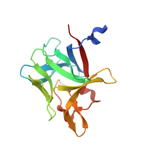
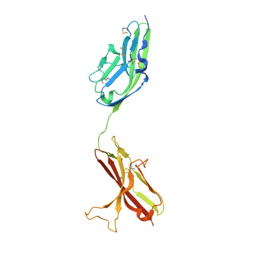
    Binding specificity between fibroblast growth factors (FGFs) and their receptors (FGFRs) is essential for mammalian development and is regulated primarily by two alternatively spliced exons, IIIb ("b") and IIIc ("c"), that encode the second half of Ig-like domain 3 (D3) of FGFRs. FGF7 and FGF10 activate only the b isoform of FGFR2 (FGFR2b). Here, we report the crystal structure of the ligand-binding portion of FGFR2b bound to FGF10. Unique contacts between divergent regions in FGF10 and two b-specific loops in D3 reveal the structural basis by which alternative splicing provides FGF10-FGFR2b specificity. Structure-based mutagenesis of FGF10 confirms the importance of the observed contacts for FGF10 biological activity. Interestingly, FGF10 binding induces a previously unobserved rotation of receptor Ig domain 2 (D2) to introduce specific contacts with FGF10. Hence, both D2 and D3 of FGFR2b contribute to the exceptional specificity between FGF10 and FGFR2b. We propose that ligand-induced conformational change in FGFRs may also play an important role in determining specificity for other FGF-FGFR complexes.


  • Organizational Affiliation

    Department of Pharmacology, New York University School of Medicine, New York, NY 10016, USA.


Macromolecules
Find similar proteins by:  (by identity cutoff)  |  3D Structure
Entity ID: 1
MoleculeChains Sequence LengthOrganismDetailsImage
Fibroblast growth factor-10145Homo sapiensMutation(s): 0 
Gene Names: FGF10
UniProt & NIH Common Fund Data Resources
Find proteins for O15520 (Homo sapiens)
Explore O15520 
Go to UniProtKB:  O15520
PHAROS:  O15520
GTEx:  ENSG00000070193 
Entity Groups  
Sequence Clusters30% Identity50% Identity70% Identity90% Identity95% Identity100% Identity
UniProt GroupO15520
Sequence Annotations
Expand
  • Reference Sequence
Find similar proteins by:  (by identity cutoff)  |  3D Structure
Entity ID: 2
MoleculeChains Sequence LengthOrganismDetailsImage
fibroblast growth factor receptor 2 isoform 2230Homo sapiensMutation(s): 4 
UniProt & NIH Common Fund Data Resources
Find proteins for P21802 (Homo sapiens)
Explore P21802 
Go to UniProtKB:  P21802
PHAROS:  P21802
GTEx:  ENSG00000066468 
Entity Groups  
Sequence Clusters30% Identity50% Identity70% Identity90% Identity95% Identity100% Identity
UniProt GroupP21802
Sequence Annotations
Expand
  • Reference Sequence
Small Molecules
Ligands 2 Unique
IDChains Name / Formula / InChI Key2D Diagram3D Interactions
15P
Query on 15P

Download Ideal Coordinates CCD File 
E [auth B]POLYETHYLENE GLYCOL (N=34)
C69 H140 O35
VUYXVWGKCKTUMF-UHFFFAOYSA-N
SO4
Query on SO4

Download Ideal Coordinates CCD File 
C [auth A],
D [auth A]
SULFATE ION
O4 S
QAOWNCQODCNURD-UHFFFAOYSA-L
Modified Residues  1 Unique
IDChains TypeFormula2D DiagramParent
MSE
Query on MSE
B
L-PEPTIDE LINKINGC5 H11 N O2 SeMET
Experimental Data & Validation

Experimental Data

  • Method: X-RAY DIFFRACTION
  • Resolution: 2.90 Å
  • R-Value Free: 0.288 
  • R-Value Work: 0.239 
  • R-Value Observed: 0.232 
  • Space Group: P 64 2 2
Unit Cell:
Length ( Å )Angle ( ˚ )
a = 113.93α = 90
b = 113.93β = 90
c = 164.852γ = 120
Software Package:
Software NamePurpose
CNSrefinement
DENZOdata reduction
SCALEPACKdata scaling
CNSphasing

Structure Validation

View Full Validation Report


Entry History 

Deposition Data

Revision History  (Full details and data files)

  • Version 1.0: 2003-02-11
    Type: Initial release
  • Version 1.1: 2008-04-29
    Changes: Version format compliance
  • Version 1.2: 2011-07-13
    Changes: Version format compliance